. 2022 Jan:175:105995.
doi: 10.1016/j.phrs.2021.105995. Epub 2021 Nov 21.

Identification of Piezo1 channels in perivascular adipose tissue (PVAT) and their potential role in vascular function

Affiliations

Identification of Piezo1 channels in perivascular adipose tissue (PVAT) and their potential role in vascular function

Taylor R Miron et al. Pharmacol Res. 2022 Jan.

Abstract

The vasculature constantly experiences distension/pressure exerted by blood flow and responds to maintain homeostasis. We hypothesized that activation of the stretch sensitive, non-selective cation channel Piezo1 would directly increase vascular contraction in a way that might be modified by perivascular adipose tissue (PVAT). The presence and function of Piezo1 was investigated by RT-PCR, immunohistochemistry, and isolated tissue bath contractility. Superior and mesenteric resistance arteries, aortae, and their PVATs from male Sprague Dawley rats were used. Piezo1 mRNA was detected in aortic vessels, aortic PVAT, mesenteric vessels, and mesenteric PVAT. Both adipocytes and stromal vascular fraction of mesenteric PVAT expressed Piezo1 mRNA. In PVAT, expression of Piezo1 mRNA was greater in magnitude than that of Piezo2, transient receptor potential cation channel, subfamily V, member 4 (TRPV4), anoctamin 1, calcium activated chloride channel (TMEM16), and Pannexin1 (Panx1). Piezo1 protein was present in endothelium and PVAT of rat aortic and in PVAT of mesenteric artery. The Piezo1 agonists Yoda1 and Jedi2 (1 nM - 10 µM) did not stimulate aortic contraction [max < 10% phenylephrine (PE) 10 µM contraction] or relaxation in tissues + or -PVAT. Depolarizing the aorta by modestly elevated extracellular K+ did not unmask aortic contraction to Yoda1 (max <10% PE 10 µM contraction). Finally, the Piezo1 antagonist Dooku1 did not modify PE-induced aorta contraction + or -PVAT. Surprisingly, Dooku1 directly caused aortic contraction in the absence (Dooku1 =26 ± 11; Vehicle = 11 ± 11%PE contraction) but not in the presence of PVAT (Dooku1 = 2 ± 1; Vehicle = 8 ± 5% PE contraction). Thus, Piezo1 is present and functional in the isolated rat aorta but does not serve direct vascular contraction with or without PVAT. We reaffirmed the isolated mouse aorta relaxation to Yoda1, indicating a species difference in Piezo1 activity between mouse and rat.

Keywords: Acetylcholine (PubChem CID: 6060); Dooku1 (PubChem CID: 137321150); Endothelium; Jedi2 (PubChem CID: 2796026); PVAT; Phenylephrine (PubChem CID: 6041); Piezo; Vascular; Yoda1 (PubChem CID: 2746822).


Conflict of interest statement

Declaration of interest

The authors declare no conflict of interest.

Figures
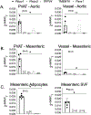
Fig. 1.
Fig. 1.
Piezo1 mRNA is present in vessels and PVAT of the male Sprague Dawley rat aorta (A) and mesenteric vessel (B) as well as in the adipocytes and SVF of the mesenteric PVAT(C). Piezo1, Piezo2, TRPV4, TMEM16, and Panx1 mRNA were measured using RT-PCR and beta actin as a calibrator control. Bars are mean+SEM with scattered individual points for an N = 4–5. Stat test was a one-way ANOVA, *p < 0.05.
Fig. 2.
Fig. 2.
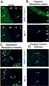
Detection of Piezo1 in distinct layers (endothelium [E], media [M], and PVAT [P]) of isolated rat thoracic aorta (A), superior mesenteric artery (B), mesenteric resistance vessels (artery [A] and vein [V]) (C), and positive control rat kidney (D). L = lumen. Top panel for each set of images shows DAPI as a nuclei marker and FITC indicating Piezo1 signal, while bottom panel shows tissue without primary antibody (no 1°). White arrows indicate staining of interest. Images are representative of N = 5 taken at 20x with 100 μm scale bars bottom right.
Fig. 3.
Fig. 3.
Lack of immunohistochemical staining for Piezo2 in isolated rat thoracic aorta (A) and mesenteric resistance artery (B). Positive control of rat skin (C). L = lumen, E = endothelial layer, M = medial layer, P = PVAT layer, A = artery, and V = vein. Top panel for each set of images shows DAPI as a nuclei marker and FITC indicating Piezo2 signal, while bottom panel shows tissue without primary antibody (no 1°). White arrows indicate staining of potential interest. Images are representative of N = 4 taken at 20x with 100 μm scale bar bottom right.
Fig. 4.
Fig. 4.
(A) Representative tracing of contractile response to cumulative additions of the Piezo1 agonist Yoda1 to thoracic aorta ring with PVAT. Piezo1 agonists did not cause a concentration dependent contraction. Quantification of multiple experiments using Yoda1 (B) and Jedi2 (C) as agonists (grey) vs Vehicle (white, same vehicle for B and C). (A) Arrows indicate each cumulative step with PE maximal contraction at the end. (B, C) Symbols represent mean±SEM for N reported, −P = without PVAT (open) +P = with PVAT (filled).
Fig. 5.
Fig. 5.
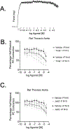
(A) Representative tracing of relaxant response to cumulative additions of the Piezo1 agonist Yoda1 in half-maximally PE contracted thoracic aorta ring with PVAT. Neither the Piezo1 agonist Yoda1 (B) nor Jedi2 (C) (grey) caused concentration dependent relaxation vs Vehicle (black). (A) Arrows indicate each cumulative step. (B, C) Symbols represent mean±SEM for N reported, −P = without PVAT (open) +P = with PVAT (filled).
Fig. 6.
Fig. 6.
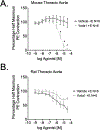
Piezo1 agonist Yoda1 induced relaxation of the longitudinal strip of thoracic aorta with endothelium in mouse (A) but not rat (B). (A,B) +E = Endothelium intact (ACh caused >40% PE relaxation), PVAT removed, thoracic aorta in strips. Points shown as mean±SEM for N reported. Star is significance from two-way ANOVA p < 0.05.
Fig. 7.
Fig. 7.
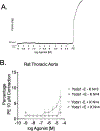
(A) Isometric tracing for Yoda1 CRC representing (B) quantification of Piezo1 agonist Yoda1 in rat thoracic aorta rings without PVAT, with and without endothelium (+E [78.22 ± 1.63% relaxation to ACh], −E [0.25 ± 0.57% relaxation to ACh], filled, open respectively) and without potassium (−K, circles) and with potassium (+K, triangles). (A) Arrows indicate each cumulative step with PE maximal contraction at the end. (B) Symbols represent mean±SEM for N reported.
Fig. 8.
Fig. 8.
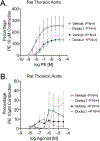
(A) PE concentration dependent contraction in isolated thoracic aorta in the presence of vehicle (DMSO, black) or Dooku1 (10 μM, grey) with or without PVAT (filled or open symbols, respectively). (B) Dooku1 concentration response curve (grey) vs vehicle (DMSO, black) in isolated thoracic aorta ring with or without PVAT (filled or open symbols, respectively). Symbols shown represent mean±SEM for N reported.
Fig. 9.
Fig. 9.
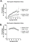
Effect of vehicle (DMSO, black) or Piezo1 agonist Yoda1 (grey) on rat superior mesenteric artery contraction (A) or relaxation (B; half-maximally contracted with PE) with or without PVAT (filled or open symbols, respectively). Points represent mean±SEM for N reported.

References

    1. Coste B, Mathur J, Schmidt M, Earley TJ, Ranade S, Petrus MJ, Dubin AE, Patapoutian A, Piezo1 and Piezo2 are essential components of distinct mechanically activated cation channels, Science 330 (6000) (2010) 55–60. - PMC - PubMed
    1. Moroni M, Servin-Vences MR, Fleischer R, Sánchez-Carranza O, Lewin GR, Voltage gating of mechanosensitive PIEZO channels, Nat. Commun. 9 (1) (2018) 1096. - PMC - PubMed
    1. Retailleau K, Duprat F, Arhatte M, Ranade SS, Peyronnet R, Martins JR, Jodar M, Moro C, Offermanns S, Feng Y, Demolombe S, Patel A, Honoré E, Piezo1 in smooth muscle cells is involved in hypertension-dependent arterial remodeling, Cell Rep. 13 (6) (2015) 1161–1171. - PubMed
    1. Nourse JL, Pathak MM, How cells channel their stress: interplay between Piezo1 and the cytoskeleton, Semin Cell Dev. Biol. 71 (2017) 3–12. - PMC - PubMed
    1. Ridone P, Vassalli M, Martinac B, Piezo1 mechanosensitive channels: what are they and why are they important, Biophys. Rev. 11 (5) (2019) 795–805. - PMC - PubMed

Publication types

LinkOut - more resources