. 2021 Dec:48:102192.
doi: 10.1016/j.redox.2021.102192. Epub 2021 Nov 18.

Compensatory role of endogenous sulfur dioxide in nitric oxide deficiency-induced hypertension

Affiliations

Compensatory role of endogenous sulfur dioxide in nitric oxide deficiency-induced hypertension

Yunjia Song et al. Redox Biol. 2021 Dec.

Abstract

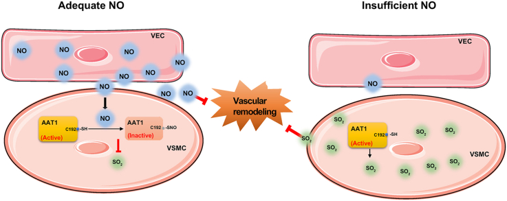
Objective: This study aimed to determine the communicational pattern of gaseous signaling molecules sulfur dioxide (SO2) and nitric oxide (NO) between vascular endothelial cells (VECs) and vascular smooth muscle cells (VSMCs), and elucidate the compensatory role and significance of endogenous SO2 in the development of hypertension due to NO deficiency.

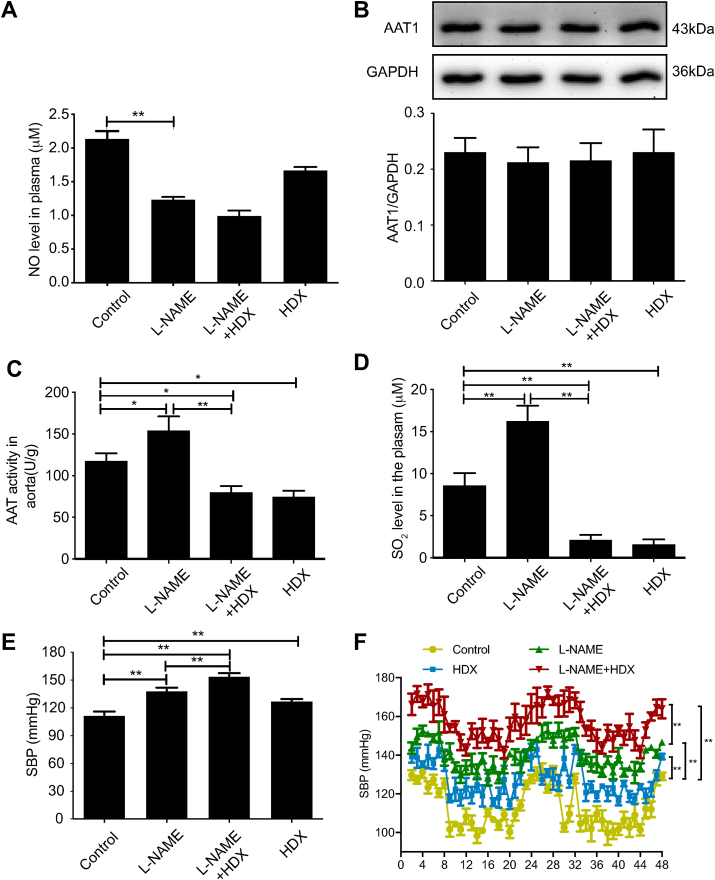
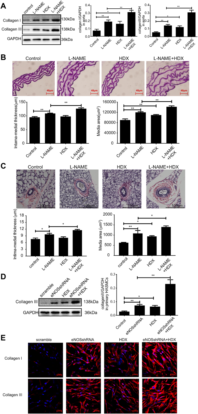
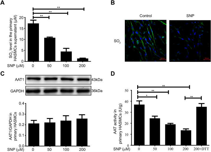
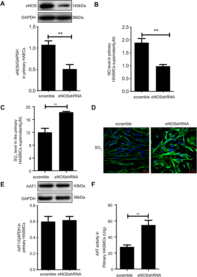
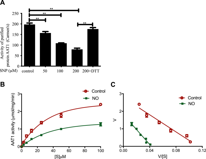
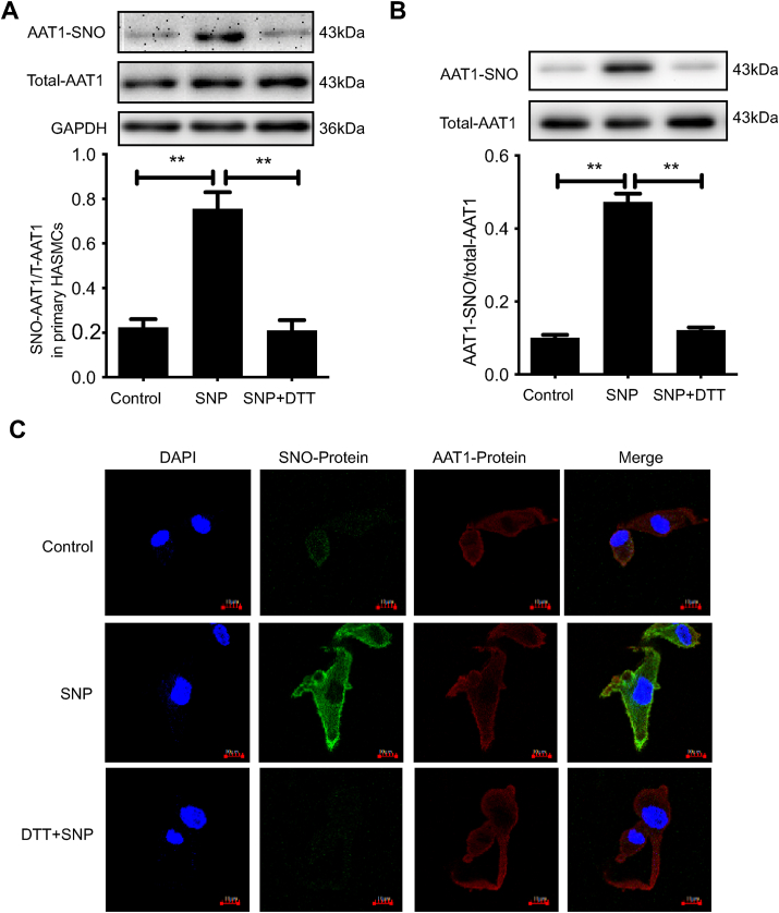
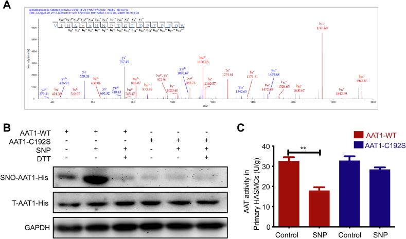
Approach and results: Blood pressure was monitored by the tail-cuff and implantable physiological signal telemetry in L-nitro-arginine methyl ester (l-NAME)-induced hypertensive mice, and structural alterations of mouse aortic vessels were detected by the elastic fiber staining method. l-NAME-treated mice showed decreased plasma NO levels, increased SO2 levels, vascular remodeling, and increased blood pressure, and application of l-aspartate-β-hydroxamate, which inhibits SO2 production, further aggravated vascular structural remodeling and increased blood pressure. Moreover, in a co-culture system of HAECs and HASMCs, NO from HAECs did not influence aspartate aminotransferase (AAT)1 protein expression but decreased AAT1 activity in HASMCs, thereby resulting in the inhibition of endogenous SO2 production. Furthermore, NO promoted S-nitrosylation of AAT1 protein in HASMCs and purified AAT1 protein. Liquid chromatography with tandem mass spectrometry showed that the Cys192 site of AAT1 purified protein was modified by S-nitrosylation. In contrast, dithiothreitol or C192S mutations in HASMCs blocked NO-induced AAT1 S-nitrosylation and restored AAT1 enzyme activity.

Conclusion: Endothelium-derived NO inhibits AAT activity by nitrosylating AAT1 at the Cys192 site and reduces SO2 production in HASMCs. Our findings suggest that SO2 acts as a compensatory defense system to antagonize vascular structural remodeling and hypertension when the endogenous NO pathway is disturbed.

Keywords: Aspartate aminotransferase; Endothelial cells; Remodeling; S-Nitrosylation; Sulfur dioxide.


Conflict of interest statement

The authors declare that they have no conflict of interest.

Figures

Image 1
Graphical abstract
Fig. 1
Fig. 1
Endogenous SO2 pathway compensates disturbed endogenous NO generation to protect against hypertension. A, Colorimetric assay of plasma NO content in mice (n = 8). B, Western blotting analysis to detect the expression of AAT1 protein in mouse aortic tissues (n = 8). C, Colorimetric assay of AAT activity in mouse aortic tissues (n = 8). D, Colorimetric assay to determine SO2 in mouse plasma (n = 8). E, Blood pressure measurement in mice by the tail-cuff method (n = 8). F, Blood pressure monitoring in mice by implantable telemetry (n = 3). Data are expressed as mean ± SEM. *P < 0.05, **P < 0.01. AAT, aspartate aminotransferase; NO, nitric oxide; SO2, sulfur dioxide.
Fig. 2
Fig. 2
Endogenous SO2 pathway compensates disturbed endogenous NO generation to protect against vascular structural remodeling A, Western blot analysis to detect collagen I and III protein expression in mouse aortic tissues (n = 8). B, Elastic fiber staining method to detect the median area and thickness in the mouse aortic section (n = 8). Scale bar, 40 μm. C, Verhoeff's-Van-Gieson staining of arterioles in mouse brown fat tissues (n = 8). Scale bar, 100 μm. D, Primary HAECs were transfected with scramble or eNOS shRNA lentivirus and co-cultured with primary HASMCs. Primary HASMCs were incubated with 100 μM HDX in the culture supernatant for 24 h. Western blotting analysis was performed to detect the protein expression of collagen III in primary HASMCs in the co-culture experiment (n = 10). E, Primary HAECs were transfected with scramble or eNOS shRNA lentivirus and co-cultured with primary HASMCs. Primary HASMCs were incubated with 100 μM HDX in the culture supernatant for 24 h. Collagen I and III protein expression in primary HASMCs was detected by immunofluorescence in the co-culture experiment (n = 9). The nucleus and collagen I/III are indicated by blue and red fluorescence, respectively. Scale bar, 30 μm **P < 0.01. HDX, l-aspartate-β-hydroxamate; NO, nitric oxide; SO2, sulfur dioxide. (For interpretation of the references to color in this figure legend, the reader is referred to the Web version of this article.)
Fig. 3
Fig. 3
NO donor inhibits endogenous SO2 production by suppressing AAT1 activity in primary HASMCs. A, Primary HASMCs were treated by varying concentrations of SNP (0, 50, 100, or 200 μM) for 2 h. SO2 content in the supernatant in primary HASMCs was detected by HPLC. B, Primary HASMCs were treated with 100 μM SNP for 2 h, and endogenous SO2 content in primary HASMCs was detected by the SO2 fluorescence probe method. The nucleus is indicated by blue fluorescence, and SO2 by green fluorescence. Scale bar, 30 μm. C, Primary HASMCs were treated with various concentrations of SNP (0, 50, 100, or 200 μM) for 2 h, and AAT1 protein expression was detected in primary HASMCs by western blotting analysis. D, Primary HASMCs were treated with various concentrations of SNP (0, 50, 100, or 200 μM) for 2 h or with 200 μM SNP for 1 h and then DTT (100 μM) for 1 h. Colorimetric assay was used to detect AAT activity in primary HASMCs. *P < 0.05, **P < 0.01. n = 9–12. Data are expressed as mean ± SEM. NO, nitric oxide; SNP, sodium nitroprusside; SO2, sulfur dioxide. (For interpretation of the references to color in this figure legend, the reader is referred to the Web version of this article.)
Fig. 4
Fig. 4
eNOS knockdown facilitates endogenous SO2 production by promoting AAT1 activity in primary HASMCs. Primary HAECs were transfected with scramble or eNOS shRNA lentivirus, and the stably transfected lentiviral HAECs were co-cultured with HASMCs. A, Western blotting analysis to detect eNOS protein expression in primary HAECs in the co-culture system. B, A colorimetric assay to detect the supernatant NO content in the primary HAECs in the co-culture system. C, HPLC method for determining SO2 in the supernatant in primary HASMCs in the co-culture system. D, SO2 fluorescence probe to detect endogenous SO2 in primary HASMCs in the co-culture system. The nucleus is indicated by blue fluorescence, and SO2 by green fluorescence. Scale bar, 20 μm. E, Western blotting analysis to detect AAT1 protein expression in primary HASMCs in the co-culture system. F, Colorimetric assay to detect AAT activity in primary HASMCs in the co-culture system. **P < 0.01. n = 9–12. Data are expressed as mean ± SEM. HDX, l-aspartate-β-hydroxamate; NO, nitric oxide; SO2, sulfur dioxide. (For interpretation of the references to color in this figure legend, the reader is referred to the Web version of this article.)
Fig. 5
Fig. 5
NO directly inhibits the activity of AAT1 purified protein and affects kinetic characteristics of the AAT1 enzyme. A, Purified AAT1 proteins were treated with various concentrations of SNP (0, 50, 100, or 200 μM) for 2 h or with SNP (200 μM) for 1 h and then DTT (100 μM) for 1 h. The purified protein AAT activity was detected by colorimetric assay (n = 6). B and C, 100 μM SNP or ddH2O treatment for 30 min at 37°C. The Eadie-Hofstee plot graphing was used to determine Vmax and Km values of purified AAT1 protein (n = 6). **P < 0.01. Data are expressed as mean ± SEM. "V" means the reaction velocity. "[S](uM)" means the substrate concentration. AAT, aspartate aminotransferase; DTT, dithiothreitol; NO, nitric oxide; SNP, sodium nitroprusside.
Fig. 6
Fig. 6
NO inhibits AAT1 activity through S-nitrosylation. A, Primary HASMCs were treated with 100 μM of SNP for 2 h or with SNP (100 μM) for 1 h and then DTT (100 μM) for 1 h. The BSA method was used to detect nitrosylated AAT1 in primary HASMCs. B, Purified proteins were treated with 100 μM of SNP for 2 h or with SNP (100 μM) for 1 h and then DTT (100 μM) for 1 h. The BSA assay was used to detect S-nitrosylation of purified AAT1 protein. C, Primary HASMCs were treated with 100 μM of SNP for 2 h or with SNP (100 μM) for 1 h and then DTT (100 μM) for 1 h. In situ detection of AAT1 in primary HASMCs was performed by the fluorescent probe method. The nucleus is indicated by blue fluorescence, S-nitrosylation-modified protein by green fluorescence, AAT1 protein by red fluorescence, and S-nitrosylation-modified AAT1 protein by yellow fluorescence. Scale bar, 10 μm. n = 9–13.**P < 0.01. Data are expressed as mean ± SEM. AAT, aspartate aminotransferase; DTT, dithiothreitol; NO, nitric oxide; SNP, sodium nitroprusside. (For interpretation of the references to color in this figure legend, the reader is referred to the Web version of this article.)
Fig. 7
Fig. 7
NO nitrosylates AAT1 at Cys192. A, Effect of NO on the S-nitrosylation modified site of purified AAT1 protein. B, Primary HASMCs were treated with 100 μM SNP for 2 h or with SNP (100 μM) for 1 h and then DTT (100 μM) for 1 h after transfection with plasmid. The BSA method was used to detect S-nitrosylation modification of AAT1 protein Cys192 in primary HASMCs (n = 10). C, Primary HASMCs were treated with 100 μM of SNP for 2 h after transfection with plasmids, and AAT1-WT and AAT1-C192S activities in primary HASMCs were detected by colorimetric assay (n = 12). **P < 0.01. Data are expressed as mean ± SEM. AAT, aspartate aminotransferase; DTT, dithiothreitol; NO, nitric oxide; SNP, sodium nitroprusside.

References

    1. Kearney P.M., Whelton M., Reynolds K., Muntner P., Whelton P.K., He J. Global burden of hypertension: analysis of worldwide data. Lancet. 2005;365:217–223. - PubMed
    1. Oparil S., Acelajado M.C., Bakris G.L., Berlowitz D.R., Cifkova R., Dominiczak A.F., Grassi G., Jordan J., Poulter N.R., Rodgers A., Whelton P.K. Hypertension. Nat. Rev. Dis. Primers. 2018;4:18014. - PMC - PubMed
    1. Rapoport R.M., Draznin M.B., Murad F. Endothelium-dependent relaxation in rat aorta may be mediated through cyclic GMP-dependent protein phosphorylation. Nature. 1983;306:174–176. - PubMed
    1. Forstermann U., Mulsch A., Bohme E., Busse R. Stimulation of soluble guanylate cyclase by an acetylcholine-induced endothelium-derived factor from rabbit and canine arteries. Circ. Res. 1986;58:531–538. - PubMed
    1. Ignarro L.J., Harbison R.G., Wood K.S., Kadowitz P.J. Activation of purified soluble guanylate cyclase by endothelium-derived relaxing factor from intrapulmonary artery and vein: stimulation by acetylcholine, bradykinin and arachidonic acid. J. Pharmacol. Exp. Therapeut. 1986;237:893–900. - PubMed

LinkOut - more resources