. 2021 Nov 24;19(1):474.
doi: 10.1186/s12967-021-03126-4.

High efficiency closed-system gene transfer using automated spinoculation

Affiliations

High efficiency closed-system gene transfer using automated spinoculation

Victoria Ann Remley et al. J Transl Med. .

Abstract

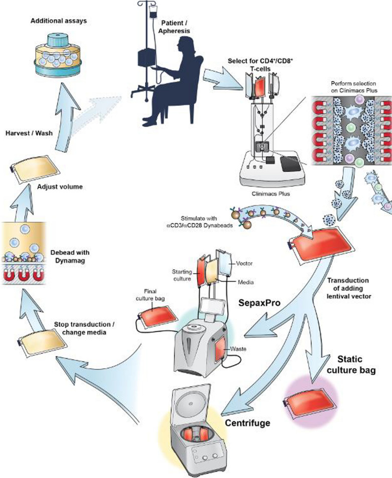
Background: Gene transfer is an important tool for cellular therapies. Lentiviral vectors are most effectively transferred into lymphocytes or hematopoietic progenitor cells using spinoculation. To enable cGMP (current Good Manufacturing Practice)-compliant cell therapy production, we developed and compared a closed-system spinoculation method that uses cell culture bags, and an automated closed system spinoculation method to decrease technician hands on time and reduce the likelihood for microbial contamination.

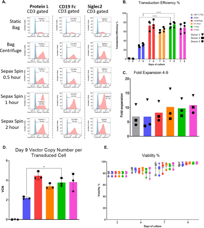
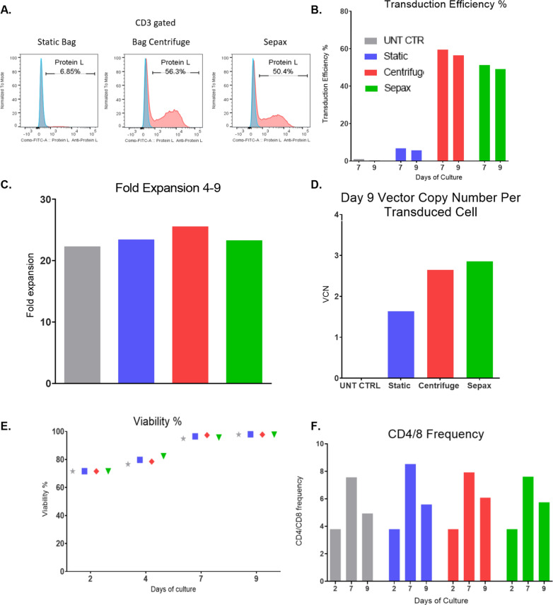
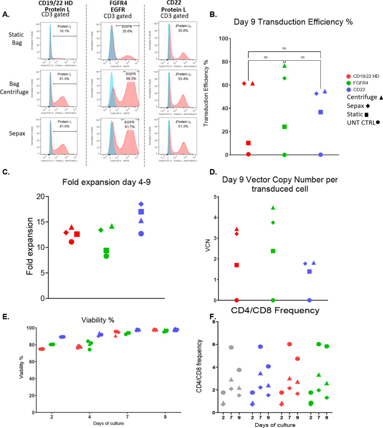
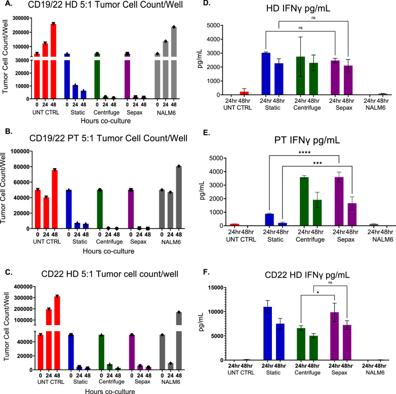
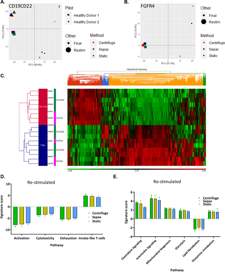
Methods: Sepax spinoculation, bag spinoculation, and static bag transduction without spinoculation were compared for lentiviral gene transfer in lymphocytes collected by apheresis. The lymphocytes were transduced once and cultured for 9 days. The lentiviral vectors tested encoded a CD19/CD22 Bispecific Chimeric Antigen Receptor (CAR), a FGFR4-CAR, or a CD22-CAR. Sepax spinoculation times were evaluated by testing against bag spinoculation and static transduction to optimize the Sepax spin time. The Sepax spinoculation was then used to test the transduction of different CAR vectors. The performance of the process using healthy donor and a patient sample was evaluated. Functional assessment was performed of the CD19/22 and CD22 CAR T-cells using killing assays against the NALM6 tumor cell line and cytokine secretion analysis. Finally, gene expression of the transduced T-cells was examined to determine if there were any major changes that may have occurred as a result of the spinoculation process.

Results: The process of spinoculation lead to significant enhancement in gene transfer. Sepax spinoculation using a 1-h spin time showed comparable transduction efficiency to the bag spinoculation, and much greater than the static bag transduction method (83.4%, 72.8%, 35.7% n = 3). The performance of three different methods were consistent for all lentiviral vectors tested and no significant difference was observed when using starting cells from healthy donor versus a patient sample. Sepax spinoculation does not affect the function of the CAR T-cells against tumor cells, as these cells appeared to kill target cells equally well. Spinoculation also does not appear to affect gene expression patterns that are necessary for imparting function on the cell.

Conclusions: Closed system-bag spinoculation resulted in more efficient lymphocyte gene transfer than standard bag transductions without spinoculation. This method is effective for both retroviral and lentiviral vector gene transfer in lymphocytes and may be a feasible approach for gene transfer into other cell types including hematopoietic and myeloid progenitors. Sepax spinoculation further improved upon the process by offering an automated, closed system approach that significantly decreased hands-on time while also decreasing the risk of culture bag tears and microbial contamination.

Keywords: CAR T-cell; Gene transfer; Sepax; Spinoculation.


Conflict of interest statement

The authors declare that they have no competing interests.

Figures

Fig. 1
Fig. 1
Schema of the Sepax spinoculation manufacturing process. Once the apheresis product is collected it was loaded on the Clinimacs Plus to be selected for CD4 + /CD8 + T-cells (simultaneous double positive selection). On day 0, the selected cells were incubated with anti-CD3/CD28 Dynabeads at a ratio of 3:1 (beads:cell) with a 40 IU of IL-2. These cells are incubated for 48 h before transduction. On day 2, the cells are divided into three different groups for the transduction process. For the static transduction, cells and vector were added to culture bags (Origen). Sepax spinoculation involves the cells, media, and vector be loaded to separate transfer packs and culture bags. The instrument and kit loads and washes the cells before the vector is added. The cells and vector are spun at a speed of 1000×g for 1 h. After spinoculation, cells are loaded into a culture bag (Origen). The bag spinoculation method is similar to the Sepax method, but the cells and vector are loaded into a culture bag (Origen), placed in an overwrap bag, and then loaded into the centrifuge buckets where they are spun at 1000×g for 2 h. In all methods the cells are expanded until day 9. On day 4, the Dynabeads are removed and discarded using a DynaMag CTS. On day 9 the cells are washed, assayed and cryopreserved. Cells may be thawed for functional testing, or if a clinical product, prepared for infusion in patients
Fig. 2
Fig. 2
Closed system Sepax spinoculation is an alternative to bag spinoculation. T-cells were stimulated with CD3/28 dynabeads before being divided between 6 different conditions: Bag centrifuge, 2-h Sepax, 1-h Sepax, 0.5-h Sepax, Static bag, or untransduced control (UNT CTRL). A Representative transduction efficiency from flow histograms are shown. B Cumulative flow transduction efficiency is shown. C Fold expansion as calculated from day 4–9. D The vector copy number per transduced cell at day 9. E Viability among groups on Day 0 and Day 9, with all groups being near 100% viable on day 9. Mean and SD of triplicate experiments are shown ****Indicates p ≤ 0.0001 between the static and Sepax groups
Fig. 3
Fig. 3
Sepax spinoculation is suitable for patient samples. T-cells were prepared in the same manner as in Fig. 2, however only a 2-h bag centrifuge, 1-h Sepax, Static bag, and UNT CTRL groups were used. Patient CD19/22 CAR T-cells were produced with all three methods. Representative transduction efficiencies (A) and cumulative transduction data (B) on day 9 is shown. C T-cell fold expansion from day 4–9 was calculated. D VCN was calculated using ddPCR. E Viability was tested using AOPI. F CD4/8 frequencies were obtained by flow cytometry and plotted
Fig. 4
Fig. 4
Sepax spinoculation is suitable for other lentiviral vectors. Three CAR vectors; CD19/22 Bispecific, FGFR4, and CD22 were compared to see if the Sepax spinoculation can be performed for multiple CAR productions. Representative transduction efficiencies (A) and cumulative transduction data (B) on day 9 is shown. C Fold expansion from day 4–9 was calculated. D VCN was calculated using ddPCR. E Viability was tested using AOPI. F CD4/8 frequencies were obtained by flow cytometry and plotted
Fig. 5
Fig. 5
Functional characteristics of CAR T-cells are not affected by spinoculation. Cryopreserved CD19/22 CAR T-cells from a healthy donor, patient, and CD22 CAR T-cells were thawed and co-cultured with NALM6 tumor cells. 1:1 and 5:1 ratio of CAR T-cells to tumor cells were cultured together for 24 and 48 h. AC Tumor Cell Counts per well calculated from cell counts and flow cytometry for healthy donor CD19/22 CAR T-cells (A), patient CD19/22 CAR T-cells (B), and healthy donor CD22 CAR T-cells (C). IFNγ ELISA’s were performed to measure the cytokine levels produced by the T cells in the supernatant for healthy donor CD19/22 CAR T-cells (D), patient CD19/22 CAR T-cell (E), and healthy donor CD22 CAR T-cells (F). Mean and SD of triplicate wells are shown.*Indicates p ≤ 0.05, ***Indicates p ≤ 0.001, ****Indicates p ≤ 0.0001 between the comparison of Sepax and centrifuge; and the static and Sepax groups
Fig. 6
Fig. 6
Gene expression of the final product post spinoculation vs re-stimulated CAR T cells are similar. PCA analysis PC1 vs PC2 comparing the different transduction methods and 8-day cultured samples to re-stimulated samples between two different healthy donors for CD19/22 transduction (A) and FGFR4 transduction (B). C Hierarchical clustering analysis of CD19/22 (C) and FGFR4 CAR T-cells before and after re-stimulation. Comparison of genes for Activation, Cytotoxicity, Exhaustion, ad Innate-like T cell functions were compared across each re-stimulated transduction method (D). Comparison of genes for Chemokine signaling, Interleukin Signaling, Mitochondrial biogenesis, Glycolysis, Lipid Metabolism, and Glutamine metabolism functions across each re-stimulated transduction method (E)

References

    1. Blaese RM, Culver KW, Miller AD, Carter CS, Fleisher T, Clerici M, et al. T lymphocyte-directed gene therapy for ADA- SCID: initial trial results after 4 years. Science. 1995;270(5235):475–480. doi: 10.1126/science.270.5235.475. - DOI - PubMed
    1. De Ravin SS, Wu X, Moir S, Anaya-O'Brien S, Kwatemaa N, Littel P, et al. Lentiviral hematopoietic stem cell gene therapy for X-linked severe combined immunodeficiency. Sci Transl Med. 2016;8(335):335ra57. doi: 10.1126/scitranslmed.aad8856. - DOI - PMC - PubMed
    1. Lee DW, Kochenderfer JN, Stetler-Stevenson M, Cui YK, Delbrook C, Feldman SA, et al. T cells expressing CD19 chimeric antigen receptors for acute lymphoblastic leukaemia in children and young adults: a phase 1 dose-escalation trial. Lancet. 2015;385(9967):517–528. doi: 10.1016/S0140-6736(14)61403-3. - DOI - PMC - PubMed
    1. Kochenderfer JN, Dudley ME, Kassim SH, Somerville RP, Carpenter RO, Stetler-Stevenson M, et al. Chemotherapy-refractory diffuse large B-cell lymphoma and indolent B-cell malignancies can be effectively treated with autologous T cells expressing an anti-CD19 chimeric antigen receptor. J Clin Oncol. 2015;33(6):540–549. doi: 10.1200/JCO.2014.56.2025. - DOI - PMC - PubMed
    1. Fry TJ, Shah NN, Orentas RJ, Stetler-Stevenson M, Yuan CM, Ramakrishna S, et al. CD22-targeted CAR T cells induce remission in B-ALL that is naive or resistant to CD19-targeted CAR immunotherapy. Nat Med. 2018;24(1):20–28. doi: 10.1038/nm.4441. - DOI - PMC - PubMed

Publication types

LinkOut - more resources