Review
. 2021 Nov 24;25(1):41.
doi: 10.1186/s40824-021-00243-5.

Preparation of alginate hydrogel microparticles by gelation introducing cross-linkers using droplet-based microfluidics: a review of methods

Affiliations
Review

Preparation of alginate hydrogel microparticles by gelation introducing cross-linkers using droplet-based microfluidics: a review of methods

Cheng Zhang et al. Biomater Res. .

Abstract

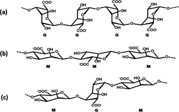
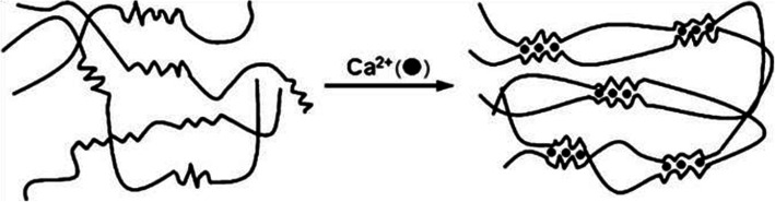
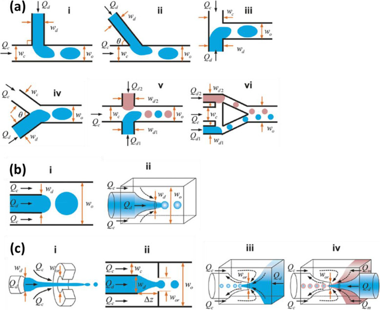
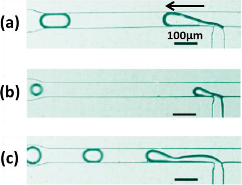
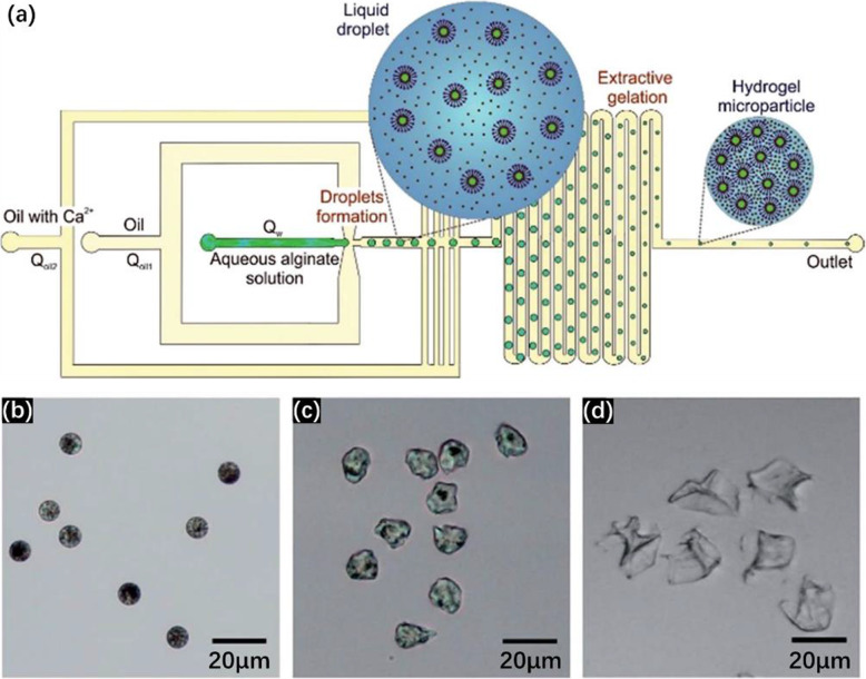
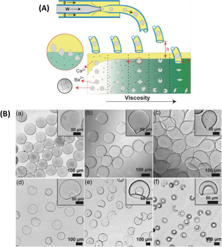
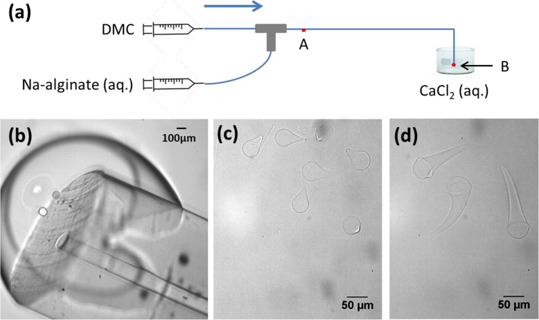
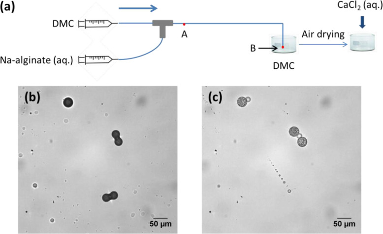
This review examines the preparation of alginate hydrogel microparticles by using droplet-based microfluidics, a technique widely employed for its ease of use and excellent control of physicochemical properties, with narrow size distribution. The gelation of alginate is realized "on-chip" and/or "off-chip", depending on where cross-linkers are introduced. Various strategies are described and compared. Microparticle properties such as size, shape, concentration, stability and mechanical properties are discussed. Finally, we consider future perspectives for the preparation of hydrogel microparticles and their potential applications.

Keywords: Alginate; Crosslinking; Droplet-based microfluidics; Hydrogel; Microparticle.


Conflict of interest statement

The authors declare no conflict of interest.

Figures

Fig. 1
Fig. 1
Chemical structures of a G-block, b M-block and c alternating G and M-blocks in alginate. Figure reprinted with permission from Reference [25]
Fig. 2
Fig. 2
Schematic illustration of the “egg-box” model describing the ionic crosslinking of alginate by calcium cations. Figure reprinted with permission from Reference [26]
Fig. 3
Fig. 3
Schematic illustrations a “cross-flowing”, b “co-flow” and c “flow-focusing” geometries for a microfluidic device. Q and w denote respectively flow rate and channel width. Subscripts d, c, o and or denote respectively dispersed fluid, continuous fluid, outlet channel and orifice. Figure reprinted with permission from Reference [65]
Fig. 4
Fig. 4
Three break-off mechanisms of droplet generation with a cross-flowing geometry: a squeezing, b dripping and c jetting. The arrow indicates the droplet flow direction. Figure reprinted with permission from Reference [57]. Copyright 2010 American Chemical Society
Fig. 5
Fig. 5
Image of connected knots formed after mixing Na-alginate and CaCl2 solutions in mineral oil with surfactant, in a PDMS-based microfluidic chip. Figure reprinted with permission from Reference [60]. Copyright 2006 American Chemical Society
Fig. 6
Fig. 6
a Schematic diagram of a PDMS-based microfluidic device using DMC as the continuous fluid in which water is partially soluble. Droplet shrinkage is observed for an initial concentration of Na-alginate of 0.5 wt%. b Micrograph of Ca-alginate hydrogel microparticles collected in an aqueous solution of CaCl2. Figure reprinted with permission from Reference [39]. Copyright 2008 American Chemical Society
Fig. 7
Fig. 7
Schematic diagram of the generation of droplets of Ca-alginate with DMC as the continuous fluid and an aqueous mixed solution of Na-alginate and CaCl2 as the dispersed fluid. The device is composed of polyether ether ketone (PEEK) junctions and Teflon-like tubing (IDEX Health and Science). The arrow indicates the flow direction
Fig. 8
Fig. 8
a Schematic diagram of the generation of Ca-alginate droplets in DMC using a cross-junction. The T-junction served to introduce DMC as a spacer to increase the distance between droplets. The device is composed of polyether ether ketone (PEEK) junctions and Teflon-like tubing (IDEX Health and Science). Micrograph of droplets observed at point A when b droplet generation was not disturbed by introducing the spacer and c when it was disturbed
Fig. 9
Fig. 9
a Schematic diagram of Ca-alginate hydrogel microparticles prepared in a PMMA based microfluidic device. b Micrographs of Ca-alginate hydrogel microparticles. Figure reprinted with permission from Reference [54]
Fig. 10
Fig. 10
a Schematic diagram of the PDMS-based microfluidic device. b Flow-focusing channel to generate alginate droplets. c Flow-focusing channel to generate CaCl2 droplets. d T-junction followed by a first circular expansion chamber. e A second circular expansion chamber. f Ca-alginate hydrogel microparticles of different shapes and sizes. Figure reprinted with permission from Reference [28]. Copyright 2006 American Chemical Society
Fig. 11
Fig. 11
a Schematic diagram of the preparation of Ca-alginate hydrogel microparticles by using CaCO3 to perform internal gelation of alginate in a PDMS-based microfluidic device. Micrograph of b droplets generated in the channel and c Ca-alginate hydrogel microparticles collected in oil. Figure reprinted with permission from Reference [61]
Fig. 12
Fig. 12
a Schematic diagram of a PDMS-based microfluidic device for the generation of droplets. b Micrograph of the two cross-junctions in the microfluidic device. c Confocal microscopic image of Ca-alginate hydrogel microparticles, some with cells encapsulated (Green fluorescence represents live cells stained by calcein AM). Figure reprinted with permission from Reference [3]
Fig. 13
Fig. 13
a Schematic diagram of a PDMS-based microfluidic device for the preparation of antigen-core alginate-shell microparticles. Inlet 1: Mineral oil with 3 wt% Span 80 and 0.2 vol% acetic acid; Inlet 2: Mineral oil with 3 wt% Span 80; Inlet 3: 2 w/v% alginate solution containing 200 mM CaCO3; Inlet 4: an antigen or protein aqueous solution. Micrographs of Ca-alginate hydrogel microparticles in b oil and c water. Figure reprinted with permission from Reference [56]
Fig. 14
Fig. 14
a Micrograph of the T-junction in a microfluidic device, where droplets of Na-alginate/Ca-EDTA were generated in oil/acetic acid. b Schematic illustration of the crosslinking process in each droplet. c Micrograph of Ca-alginate hydrogel microparticles in an aqueous medium. Figure reprinted with permission from Reference [49]
Fig. 15
Fig. 15
a Schematic diagram of the preparation of Ca-alginate hydrogel microparticles via on-chip external gelation in a PDMS-based microfluidic device. Micrographs of Ca-alginate hydrogel microparticles b in the downstream channel and c in the collecting container in soybean oil. Figure reprinted with permission from Reference [61]
Fig. 16
Fig. 16
a Schematic diagram of a glass-based microfluidic device for the preparation of Ca-alginate hydrogel microparticles, with channels modified so as to be hydrophobic. b Micrographs of hydrogel microparticles obtained with different mass fractions of the aqueous CaCl2 solution in emulsion (W). Figure reprinted with permission from Reference [29]
Fig. 17
Fig. 17
a Schematic diagram of a PDMS-based microfluidic device for the preparation of Ca-alginate hydrogel microparticles. Micrographs of microparticles with a b spherical, c slightly deformed and d collapsed morphology obtained using different experimental flow rates and calcium concentrations. Figure reprinted with permission from Reference [34]
Fig. 18
Fig. 18
A Schematic diagram of the preparation of Ca-alginate hydrogel microparticles using a microfluidic device constructed with glass capillaries, and off-chip gelation in a two-phase gelation bath. B Micrographs of Ca-alginate hydrogel microparticles of different shapes prepared under different experimental conditions. Figure reprinted with permission from Reference [20]
Fig. 19
Fig. 19
a Schematic diagram of the preparation of Ca-alginate hydrogel microparticles by off-chip external gelation without pre-gelation. Droplets were generated using a microfluidic device assembled from fluoropolymer capillaries and a T-junction. Micrographs of b the channel outlet immersed in an aqueous solution of CaCl2; Ca-alginate hydrogel microparticles prepared by collecting droplets in an aqueous solution of CaCl2 at concentrations of c 1 wt% and d 0.1 wt%
Fig. 20
Fig. 20
a Schematic diagram of a two-step preparation of Ca-alginate hydrogel microparticles using a microfluidic device constructed from fluoropolymer capillaries and junctions. Micrographs of b dried Na-alginate microparticles in air and c corresponding Ca-alginate microparticles after gelation in CaCl2 solution. Figure reprinted and adapted with permission from Reference [59]
Fig. 21
Fig. 21
Size distribution of microparticles of Na-alginate (blue) and Ca-alginate (orange) produced using droplet-based microfluidics. The curves show Gaussian fitting. Figure reprinted and adapted with permission from Reference [59]
Fig. 22
Fig. 22
SEM photographs of 2 Na-alginate microparticles (a) and (b), magnified 1000x (a1 and b1) and 5000x (a2 and b2). Na-alginate microparticles were prepared following the method mentioned in the publication [59]. Figure reprinted and adapted with permission from Reference [59]

References

    1. Agüero L, Zaldivar-Silva D, Peña L, Dias ML. Alginate microparticles as oral colon drug delivery device: a review. Carbohydr Polym. 2017;168:32–43. doi: 10.1016/j.carbpol.2017.03.033. - DOI - PubMed
    1. Ahmed EM. Hydrogel: preparation, characterization, and applications: a review. J Adv Res. 2015;6(2):105–121. doi: 10.1016/j.jare.2013.07.006. - DOI - PMC - PubMed
    1. Akbari S, Pirbodaghi T. Microfluidic encapsulation of cells in alginate particles via an improved internal gelation approach. Microfluid Nanofluid. 2014;16(4):773–777. doi: 10.1007/s10404-013-1264-z. - DOI
    1. Barbieri M, Cellini F, Cacciotti I, Peterson SD, Porfiri M. In situ temperature sensing with fluorescent chitosan-coated PNIPAAm/alginate beads. J Mater Sci. 2017;52(20):12506–12512. doi: 10.1007/s10853-017-1345-6. - DOI
    1. Baroud CN, Gallaire F, Dangla R. Dynamics of microfluidic droplets. Lab Chip. 2010;10(16):2032–2045. doi: 10.1039/c001191f. - DOI - PubMed