

Comment
doi: 10.1073/pnas.2118020118.
Mitochondria-cytoskeleton interactions in mammalian sperm revealed by cryoelectron tomography
Affiliations
- PMID: 34819381
- PMCID: PMC8640839
- DOI: 10.1073/pnas.2118020118
Item in Clipboard
Comment
Mitochondria-cytoskeleton interactions in mammalian sperm revealed by cryoelectron tomography
Proc Natl Acad Sci U S A.
.
No abstract available
Conflict of interest statement
The authors declare no competing interest.
Figures
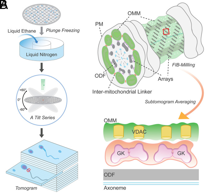
Supramolecular protein arrays at the mitochondria–cytoskeleton interface in mammalian sperm. (A) A schematic cartoon illustrating steps of obtaining a 3D reconstruction image of a plunge-frozen sperm using cryo-ET. A small volume of a suspension of sperm cells is placed onto a carbon grid and then plunge frozen in liquid ethane cooled to temperatures of ∼100 K by liquid nitrogen. The grid is then transferred into the column of an electron microscope for collection of a series of images at varying tilts. The series of tilted images is then converted to a 3D volume (tomogram) that provides a representation of the distribution of EM densities of cellular components. (B) A part of the midpiece from a mammalian sperm depicting a flagellar cross-section and an exposed mitochondria–cytoskeleton interface studded with the protein arrays. Intermitochondrial linkers are marked in orange. FIB milling can generate thin slices exposing the area of interest for subtomogram averaging. PM, plasma membrane; ODF, outer dense fiber.
Comment on
-
In-cell structures of conserved supramolecular protein arrays at the mitochondria-cytoskeleton interface in mammalian sperm.Proc Natl Acad Sci U S A. 2021 Nov 9;118(45):e2110996118. doi: 10.1073/pnas.2110996118. Proc Natl Acad Sci U S A. 2021. PMID: 34737233 Free PMC article.
References
Publication types
MeSH terms
Grants and funding
LinkOut - more resources
Full Text Sources
