. 2021 Nov 24;12(1):6815.
doi: 10.1038/s41467-021-27113-x.

Trpm5 channels encode bistability of spinal motoneurons and ensure motor control of hindlimbs in mice

Affiliations

Trpm5 channels encode bistability of spinal motoneurons and ensure motor control of hindlimbs in mice

Rémi Bos et al. Nat Commun. .

Abstract

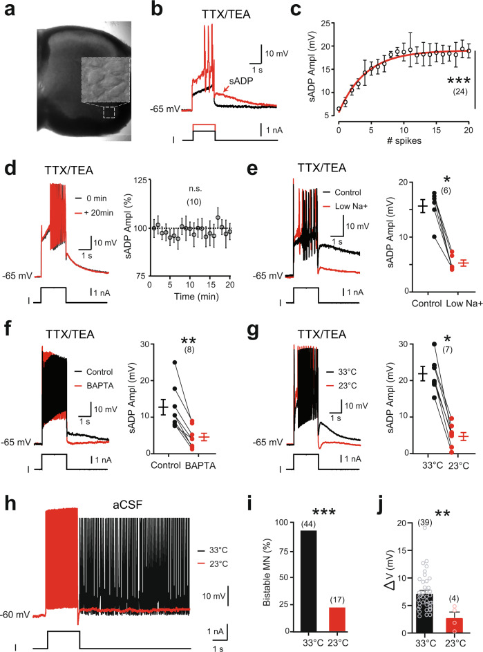
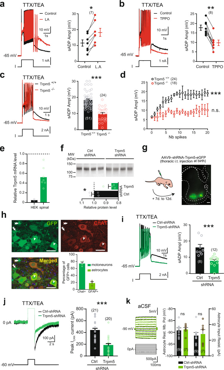
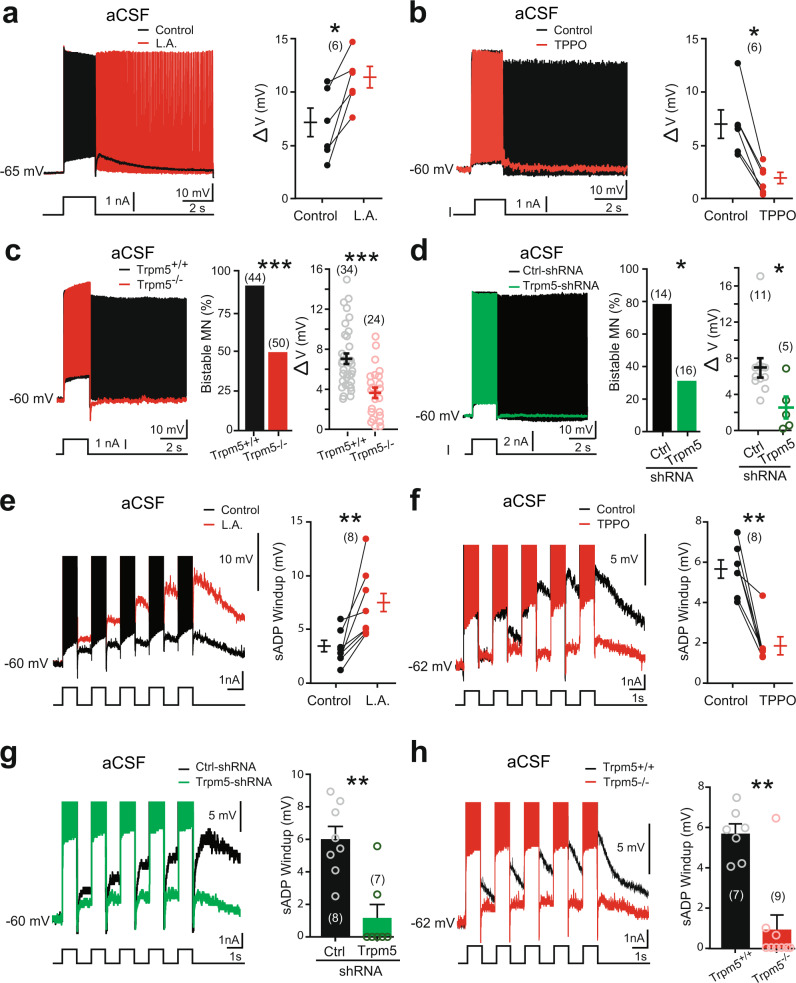
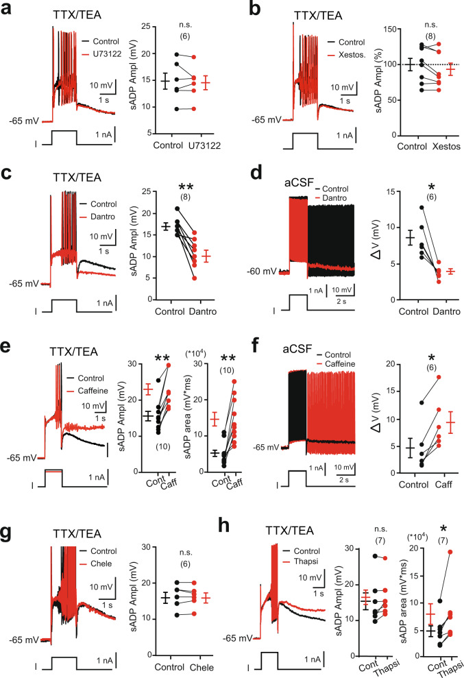
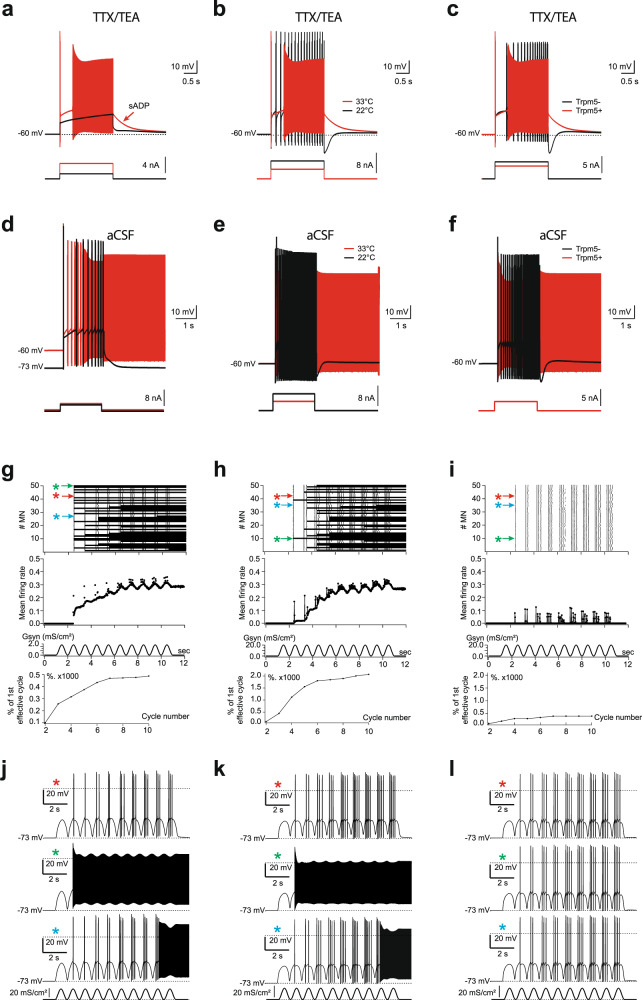
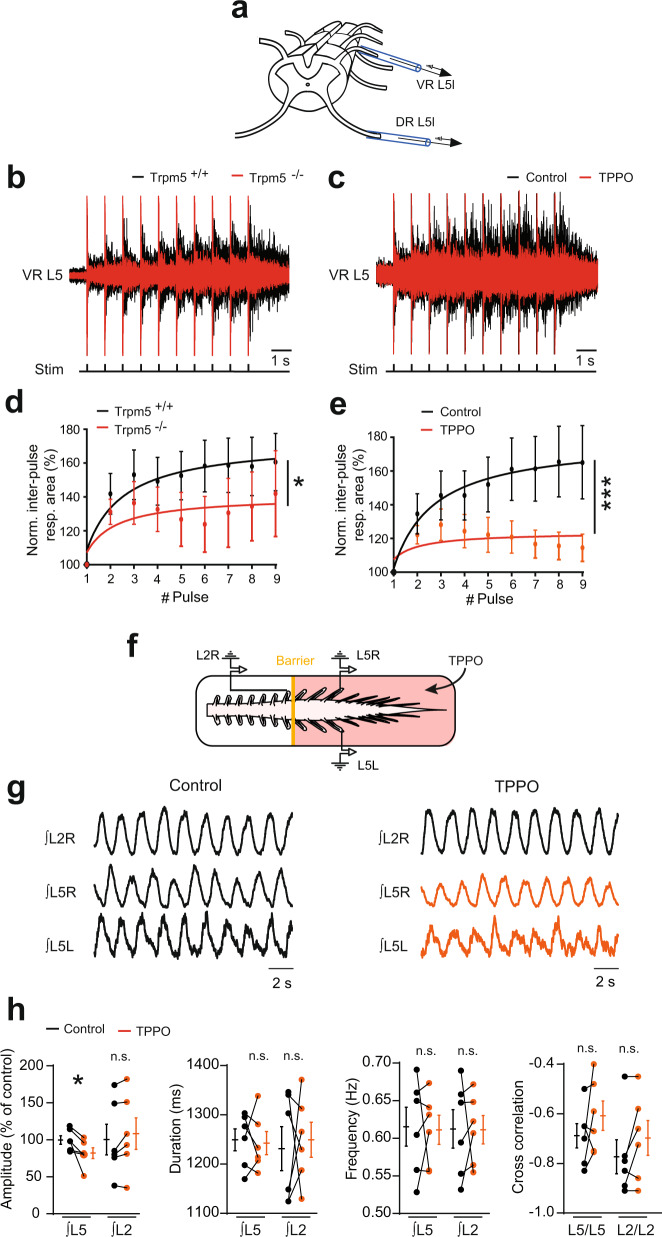
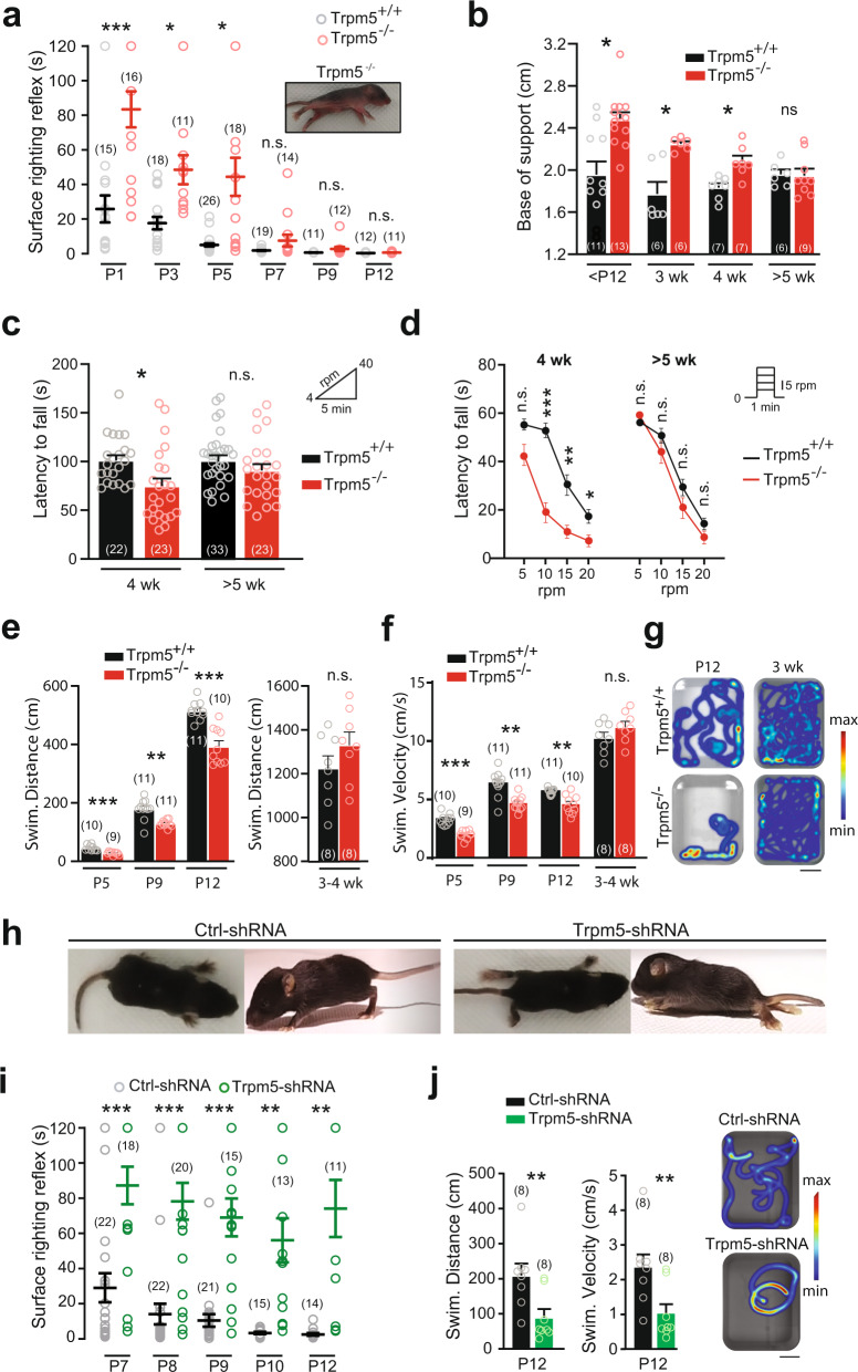
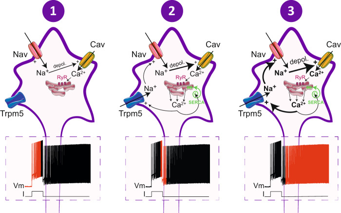
Bistable motoneurons of the spinal cord exhibit warmth-activated plateau potential driven by Na+ and triggered by a brief excitation. The thermoregulating molecular mechanisms of bistability and their role in motor functions remain unknown. Here, we identify thermosensitive Na+-permeable Trpm5 channels as the main molecular players for bistability in mouse motoneurons. Pharmacological, genetic or computational inhibition of Trpm5 occlude bistable-related properties (slow afterdepolarization, windup, plateau potentials) and reduce spinal locomotor outputs while central pattern generators for locomotion operate normally. At cellular level, Trpm5 is activated by a ryanodine-mediated Ca2+ release and turned off by Ca2+ reuptake through the sarco/endoplasmic reticulum Ca2+-ATPase (SERCA) pump. Mice in which Trpm5 is genetically silenced in most lumbar motoneurons develop hindlimb paresis and show difficulties in executing high-demanding locomotor tasks. Overall, by encoding bistability in motoneurons, Trpm5 appears indispensable for producing a postural tone in hindlimbs and amplifying the locomotor output.


Conflict of interest statement

The authors declare no competing interests.

Figures

Fig. 1
Fig. 1. Functional characterization of the thermosensitive sADP in large lumbar motoneurons from mice.
a Acute spinal cord slice from the lumbar enlargement (L4) under infrared-differential interference contrast (IR-DIC) imaged at ×4 magnification. Inset: high magnification (×40) of a motoneuronal pool readily observable in the ventral horn and patch-clamped under IR-DIC. b Superimposition of voltage traces from a motoneuron recorded under TTX and TEA in response to subthreshold (black) or suprathreshold (red) depolarizing pulses. The arrow indicates the slow after depolarization (sADP). c Relationship between the peak amplitude of the sADP and the number of spikes emerging during a 2-s depolarizing current pulse. Continuous red line is the best-fit nonlinear regression (n = 9 mice). d Mean time-course changes in peak amplitude of the sADP. Values are relative to the amplitude of the first sADP (n = 3 mice). eg Left: superimposition of voltage traces from motoneurons recorded under TTX and TEA, before and after removing [Na+]o (n = 3 mice) (e), chelating intracellular Ca2+ with BAPTA (10 mM) (n = 2 mice) (f), or decreasing temperature (n = 3 mice) (g), right: mean amplitude of the peak sADP. Each circle represents an individual motoneuron. h Superimposition of voltage traces recorded in normal aCSF (i.e., without TTX and TEA) in response to a 2-s depolarizing pulse before and after reducing the temperature of the bath from 33 to 23 °C. i, j Group mean quantification of the proportion of bistable motoneurons (i) and of the bistability range ΔV (j) as a function of temperature (n = 13 mice). Numbers in brackets indicate the numbers of recorded motoneurons. n.s., no significance; *P < 0.05; **P < 0.01; ***P < 0.001 (one-way ANOVA with multiple comparisons for c, d; two-tailed Wilcoxon paired test for eg; two-tailed Fisher test for i; two-tailed Mann–Whitney test for j). Mean ± SEM. For detailed P values, see Source data. Source data are provided as a Source data file. See also Supplementary Fig. 1.
Fig. 2
Fig. 2. The thermosensitive ICaN-mediated sADP is driven by Trpm5 channels.
ac Left: superimposition of voltage traces in motoneurons recorded under TTX/TEA from wild-type mice in response to a depolarizing pulse before and after bath-applying linoleic acid (a, L.A., 50 µM, n = 3 mice) or triphenylphosphine oxide (b, TPPO, 50 µM, n = 4 mice), or recorded in motoneurons from Trpm5−/− mice (n = 5 mice) (c), right: mean amplitude of the peak sADP. The numbers in brackets indicate the numbers of recorded motoneurons. Each circle represents an individual motoneuron. d Relationship between the peak amplitude of the sADP and the number of spikes emerging during a 2-s depolarizing current pulse in control (black, n = 9 mice) vs Trpm5−/− mice (red, n = 5 mice). e qRT-PCR analysis assessing the efficiency of the shRNA to knockdown Trpm5 mRNA in HEK-293 cell cultures (n = 2) and spinal cords (n = 7) from ~P12 mice. The expression level of the Trpm5 mRNA in cell cultures or spinal cords was normalized to scramble shRNA values with GAPDH or ACTB as internal references, respectively. Each circle represents the mean value from one cell culture or one spinal cord. f Up: Trpm5 immunoblots of lumbar segments from P12 mice intrathecally injected at birth with an adeno-associated virus (AAV9) encoding either a scramble shRNA (n = 4 mice) or a Trpm5-targeting shRNA (n = 4 mice). One mice per lane. Bottom: group mean quantification of the ~130 kDa band normalized to scramble-injected controls. g Left: schematic representation of the experimental design, right: acquisition of a transverse spinal slice (L4) from a P10 mouse intrathecally injected at birth with an AAV9 encoding Trpm5-targeting shRNA and eGFP. Scale bar = 100 μm. The experiment was repeated four independent times with similar results. h High magnification of the ventral horn showing native fluorescence of motoneurons (single arrow) transduced by AAV9 (upper left) and immunostained for choline acetyltransferase (upper right, ChAT antibody; bottom left, merged images). Some astrocytes (double arrow) were also GFP+. Histograms (bottom right): group mean quantification of the proportion of 299 motoneurons (green) and 640 astrocytes (orange) from 4 mice transfected by AAV9-shRNA-Trpm5-eGFP. Each circle represents one mouse. Scale bar = 50 μm. i, j Left: superimposition of voltage (i) or current (j) traces from GFP+ motoneurons recorded under TTX/TEA and transduced either with scramble shRNA (black, n = 6 mice) or with a Trpm5-targeting shRNA (green, n = 5 mice), right: mean amplitude of the peak sADP (i) and the peak amplitude of the ICAN current (j). k Left: superimposition of voltage traces from GFP+ astrocytes recorded in normal aCSF and transduced either with scramble shRNA (black, n = 3 mice) or with a Trpm5-targeting shRNA (green, n = 3 mice), right: mean amplitude of the astrocytic resting membrane potential (left) and the input resistance (right). The numbers in brackets indicate the numbers of recorded cells. Each circle represents an individual motoneuron or astrocyte. *P < 0.05; **P < 0.01; ***P < 0.001 (two-tailed Wilcoxon paired test for a, b; two-tailed Mann–Whitney test for c, f, ik; one-way ANOVA with multiple comparisons for d). Mean ± SEM. For detailed P values, see Source data. Source data are provided as a Source data file. See also Supplementary Figs. 2–4.
Fig. 3
Fig. 3. Bistability of motoneurons relies on Trpm5 channels.
ah Left: superimposition of voltage traces recorded in motoneurons from wild-type mice in response to a single (ad) or repetitive (1 Hz, eh) depolarizing current pulses before (black) and after (red) bath-applying linoleic acid (L.A., 30 µM) (a, e, n = 5 mice), or triphenylphosphine oxide (TPPO, 30 µM, n = 7 mice) (b, f), or recorded in motoneurons from Trpm5−/− mice (c, h, n = 7 mice) or from eGFP+ motoneurons transduced either with the scramble (black, n = 5 mice) or with a Trpm5-targeting shRNA (green, n = 7 mice) (d, g), right: group mean quantification of the proportion of bistable motoneurons and/or ΔV (ad) and of the sADP windup (eh). Numbers in brackets indicate the numbers of recorded motoneurons. Each circle represents an individual motoneuron. *P < 0.05; **P < 0.01; ***P < 0.001 (two-tailed Wilcoxon paired test for a, b, e, f; two-tailed Fisher test for c, d (middle histograms); two-tailed Mann–Whitney test for c (right), d (right), g, h). Mean ± SEM. For detailed P values, see Source data. Source data are provided as a Source data file. See also Supplementary Fig. 5.
Fig. 4
Fig. 4. Ryanodine-operated Ca2+ release activates Trpm5 to promote bistability in motoneurons.
ah Left: superimposition of voltage traces from motoneurons in response to a 2-s depolarizing current pulse recorded with (ac, e, g, h) or without (d, f) TTX/TEA before and after bath-applying U73122 (a, 10 µM, n = 2 mice), xestospongin C (b, 1–2.5 µM, n = 2 mice), dantrolene (c, d, 30 µM, n = 5 mice), caffeine (e, f, 30 µM–5 mM, n = 5 mice), chelerythrin (g, 10 µM, n = 3 mice), or thapsigargin (h, 1 µM, n = 3 mice), right: quantification of the area and/or amplitude of the sADP (ac, e, g, h) or of the ΔV (d, f) defined as the difference between the most depolarized pre-stimulus holding potential and the most hyperpolarized holding potential for which self-sustained firing can be triggered (see Supplementary Fig. 1). Numbers in brackets indicate the numbers of recorded motoneurons. n.s., no significance; *P < 0.05; **P < 0.01 (two-tailed Wilcoxon paired test). Mean ± SEM. For detailed P values, see Source data. Source data are provided as a Source data file. See also Supplementary Fig. 6.
Fig. 5
Fig. 5. Simulated motoneurons supplemented with Trpm5 channels display self-sustained spiking activity and predict a role of Trpm5 in amplifying motor outputs.
ac Superpositions of voltages generated by simulated motoneuron with diminished conductance of Na+ and K+ channels (TTX/TEA condition) in response to depolarizing 2-s pulses (bottom insets) applied at the soma initially held at −60 mV. The sADP (arrow in a) followed the spiking evoked by suprathreshold stimuli (red) in case of warm temperature (33 °C) and intact Trpm5 channels (Trpm5+) but disappeared (black) after reducing stimulation to subthreshold values (a), cooling to 22 °C (b), or blockade of Trpm5 channels (c). df Self-sustained spiking activity (red) of simulated motoneuron with intact Na+ and K+ channels triggered by a 2-s depolarizing stimuli in case of pre-holding at −60 mV, warm temperature of 33 °C, and intact Trpm5 channels (Trpm5+). Self-sustained spiking did not occur if Trpm5 channels were not sufficiently activated due to low holding (background) potential of −73 mV (d), temperature decrease to 22 °C (e), or the Trpm5 channels were totally blocked (f) although the cell remained capable of firing in response to a depolarizing pulse (black traces). gi Integrated firing activity in a population of 50 uncoupled motoneurons with randomized Trpm5 expression (normal distribution of maximum conductivity GTrpm5, mean ± s.d. = 55 ± 11 mS/cm2) generated in response to 1-Hz sinusoid synaptic excitation of the soma (g) or dendrites (h). i same as in (h), but for an ~4-fold reduced Trpm5 expression (GTrpm5 mean ± s.d. = 12.75 ± 2.0 mS/cm2). Panels top to bottom: raster plots of spiking; mean firing rate in spikes per 1 s per neuron; synaptic conductivity Gsyn associated with 0-mV reversal potential; normalized cycle-to-cycle firing rate in percentage of response to first effective cycle. Arrows in the top panel indicate scatter plots of firing of individual neurons, exemplified below in jl and marked by asterisks of the corresponding color.
Fig. 6
Fig. 6. Trpm5 channels amplify motor outputs.
a Schematic representation of the ventral spinal cord side up with the stimulating (DR L5L) and recording (VR L5L) glass electrodes. b, c Ventral root (L5) responses to 1-Hz ipsilateral dorsal root stimuli, recorded from wild-type (n = 11 mice) and Trpm5−/− (n = 7 mice) spinal cords (b) or from wild-type spinal cords before and after bath-applying triphenylphosphine oxide (TPPO, 30 µM, n = 8 mice) (c). d, e Quantification of the response as a function of the pulse number. Values are relative to the area of the initial post-stimulation response measured during the first inter-pulse interval. f Schematic representation of the whole-mount spinal cord with the recording glass electrodes from the ipsilateral (L2R, L5R) and contralateral (L5L, L5R) sides. The yellow solid line represents the Vaseline barrier separating the rostral (L2) from the caudal (L5) segments. g Ventral root recordings of NMA/5-HT-induced rhythmic activity generated before and after adding triphenylphosphine oxide (TPPO, 30 µM, n = 6 mice) to caudal lumbar segments. h Quantification of locomotor burst parameters. n.s., no significance; *P < 0.05; ***P < 0.001 (fit comparison for d, e; two-tailed Wilcoxon paired test for h). Mean ± SEM. For detailed P values, see Source data. Source data are provided as a Source data file. See also Supplementary Fig. 7.
Fig. 7
Fig. 7. Trpm5 channels ensure motor control of hindlimbs.
a Surface righting response as a function of postnatal day in wild-type (black) and Trpm5−/− (red) mice. Values represent the time spent for rotating from a supine position to a prone position on their four paws. Picture illustrates a Trpm5−/− mouse that fails to right itself within 2 min. b Quantification of the base of support between hindlimb paws during walking as a function of age in wild-type (black) and Trpm5−/− (red) mice. c Latency to fall from a rod rotating at accelerated speed (4–40 rpm) in young adult mice (4 weeks and >5 weeks old), either wild-type (black) or Trpm5−/− (red). d Latency to fall from a rod rotating at constant speed in young adult mice (4 weeks and >5 weeks old), either wild-type (black) or Trpm5−/− (red). e, f Mean swimming traveled distance (e) and velocity (f) of neonates (P5–P12) and young adult (3–4 weeks old) wild-type (black) and Trpm5−/− (red) mice during three consecutive trials. g Heatmap representation of the swimming of neonates (P12) and young adult (3 weeks old) in wild-type (top) and Trpm5−/− mice (bottom). Scale bar, 10 cm. h Top and side views of 12-day-old wild-type mice transduced either with the scramble shRNA (left) or with the Trpm5-shRNA (right). i Surface righting response during postnatal development in wild-type mice transduced either with the scramble shRNA (black) or with a Trpm5-shRNA (green). j Swimming activity of 12-day-old wild-type mice transduced either with the scramble shRNA (black) or with the Trpm5-shRNA (green) Left: Swimming distance and velocity were averaged from three consecutive swimming trials. Right: Heatmaps illustrate swimming activity. Scale bar, 10 cm. The numbers in the brackets indicate the numbers of mice. n.s., no significance; *P < 0.05; **P < 0.01; ***P < 0.001 (two-way ANOVA with Sidak’s multiple comparaisons test for ai; two-tailed Mann–Whitney test for j). Mean ± SEM. For detailed P values, see Source data. Source data are provided as a Source data file. See also Supplementary Fig. 8.
Fig. 8
Fig. 8. Overview of ionic cascades leading to bistability in spinal motoneurons.
Schematic relationships between currents underlying the different phases of the self-sustained firing mode. Trpm5 transient receptor potential cation channel subfamily M member 5, Nav voltage-gated sodium channels, Cav voltage-gated calcium channels, depol depolarization, RyR ryanodine receptors, Serca sarco/endoplasmic reticulum Ca2+-ATPase.

Similar articles

Cited by

References

    1. Bos R, et al. Kv1.2 channels promote nonlinear spiking motoneurons for powering up locomotion. Cell Rep. 2018;22:3315–3327. - PMC - PubMed
    1. Kiehn O. Plateau potentials and active integration in the ‘final common pathway’ for motor behaviour. Trends Neurosci. 1991;14:68–73. - PubMed
    1. Hultborn H. Plateau potentials and their role in regulating motoneuronal firing. Prog. Brain Res. 1999;123:39–48. - PubMed
    1. Brownstone RM, Gossard JP, Hultborn H. Voltage-dependent excitation of motoneurones from spinal locomotor centres in the cat. Exp. Brain Res. 1994;102:34–44. - PubMed
    1. Hultborn H, Brownstone RB, Toth TI, Gossard JP. Key mechanisms for setting the input-output gain across the motoneuron pool. Prog. Brain Res. 2004;143:77–95. - PubMed

Publication types

MeSH terms

LinkOut - more resources