. 2021 Dec;6(12):1575-1582.
doi: 10.1038/s41564-021-00998-6. Epub 2021 Nov 24.

Wolbachia cifB induces cytoplasmic incompatibility in the malaria mosquito vector

Affiliations

Wolbachia cifB induces cytoplasmic incompatibility in the malaria mosquito vector

Kelsey L Adams et al. Nat Microbiol. 2021 Dec.

Erratum in

Abstract

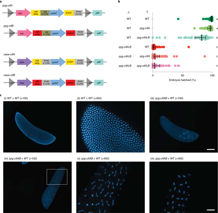
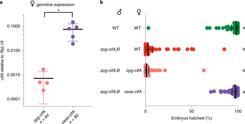
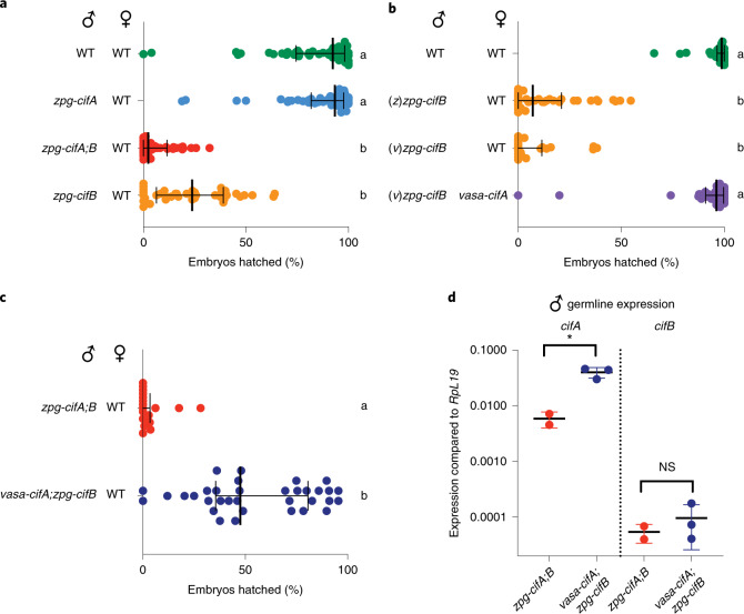
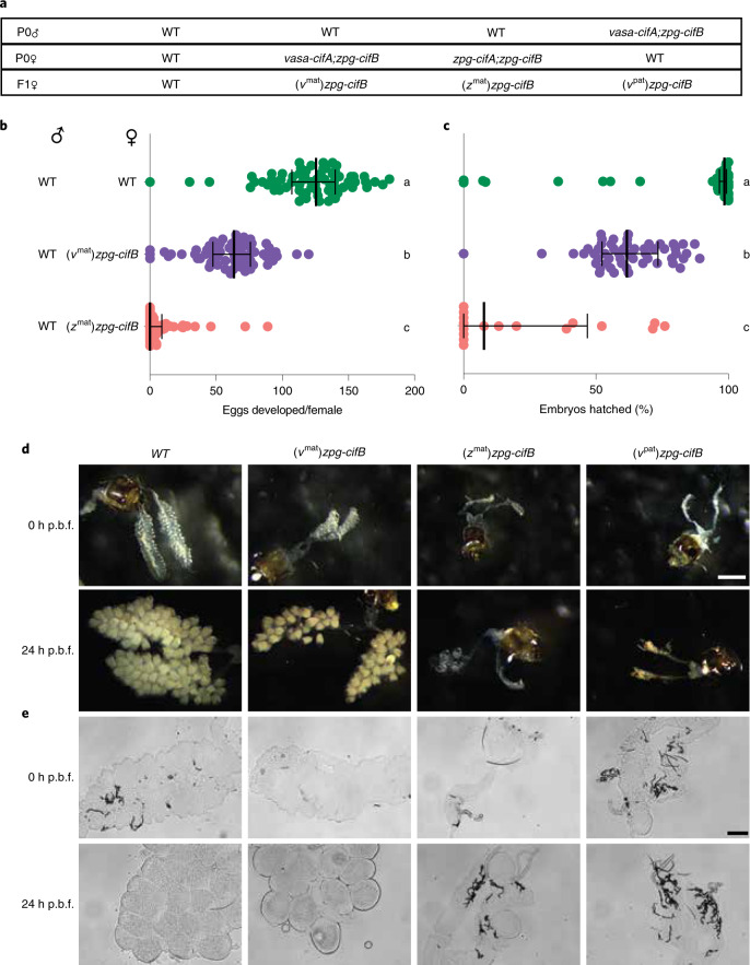
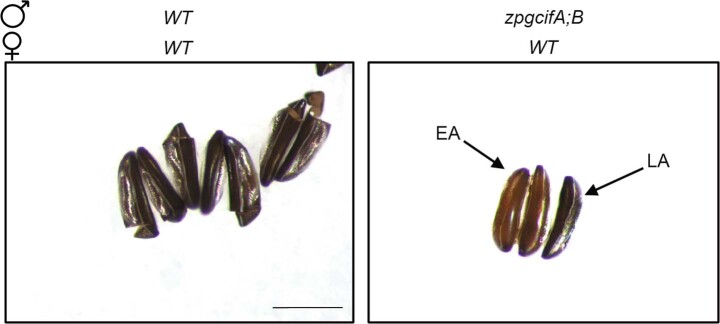
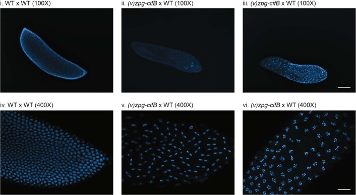
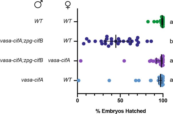
Wolbachia, a maternally inherited intracellular bacterial species, can manipulate host insect reproduction by cytoplasmic incompatibility (CI), which results in embryo lethality in crosses between infected males and uninfected females. CI is encoded by two prophage genes, cifA and cifB. Wolbachia, coupled with the sterile insect technique, has been used in field trials to control populations of the dengue vector Aedes albopictus, but CI-inducing strains are not known to infect the malaria vector Anopheles gambiae. Here we show that cifA and cifB can induce conditional sterility in the malaria vector An. gambiae. We used transgenic expression of these Wolbachia-derived genes in the An. gambiae germline to show that cifB is sufficient to cause embryonic lethality and that cifB-induced sterility is rescued by cifA expression in females. When we co-expressed cifA and cifB in male mosquitoes, the CI phenotype was attenuated. In female mosquitoes, cifB impaired fertility, which was overcome by co-expression of cifA. Our findings pave the way towards using CI to control malaria mosquito vectors.


Conflict of interest statement

The authors declare no competing interests.

Figures

Fig. 1
Fig. 1. Co-expression of cifA and cifB in male An. gambiae causes embryonic lethality in progeny.
a, Construct design of zpg-cifA, zpg-cifB, vasa-cifA and vasa-cifB. b, Males that express zpg-cifA;B produced largely inviable progeny, regardless of whether their female mate expresses zpg-cifA. Expression of zpg-cifA;B in females caused a decrease in female fertility compared with WT females, but expression of cifA alone did not (Dunn’s multiple comparisons test (two-sided), P ≤ 0.0071 for groups a vs b, P < 0.0001 for groups a vs c and groups b vs c). Median and interquartile ranges are shown. For each group (top to bottom), the n (number of broods) is as follows: 58, 52, 59, 51, 53, 62. Kruskal–Wallis test: H = 265, P < 0.0001, d.f. = 5. c, Embryos from zpg-cifA;B males crossed with WT females (or WT crosses, as controls in (i) and (ii)) were fixed and imaged with DAPI 3–4 h post oviposition, showing developmental arrest of most CI embryos during early nuclear divisions (iii), while some embryos completed multiple rounds of nuclear division but showed mitotic defects, such as chromatin bridging ((iv), with a close-up in (v)), and other chromosomal abnormalities resulting in delayed or arrested development (vi). Scale bars, 100 μm for ×100 images and 25 μm for ×400 images. Source data
Fig. 2
Fig. 2. High expression of female cifA rescues cifA;B-induced CI in An. gambiae.
a, Transcript abundance of cifA was higher in vasa-cifA females compared with zpg-cifA females relative to RpL19 (unpaired t-test (two-tailed), P = 0.0232, mean and s.d. are shown). b, The expression of vasa-cifA in females rescued infertility caused by zpg-cifA;B expression in males to WT levels, while expression of zpg-cifA in females did not (Dunn’s multiple comparisons test (two-sided), P < 0.0001 for differences between all statistical groups). Median and interquartile ranges are shown. For each group (top to bottom), the n is as follows: 51, 50, 52, 52. Kruskal–Wallis test: H = 153.1, P < 0.0001, d.f. = 3. Source data
Fig. 3
Fig. 3. Male cifB expression is sufficient to cause CI, while male cifA attenuates it.
a, zpg-cifB males caused infertility in WT females, while zpg-cifA males did not (Dunn’s multiple comparisons test (two-sided), P ≤ 0.0001 for differences between all statistical groups). Median and interquartile ranges are shown. For each group (top to bottom), the n is as follows: 55, 55, 44, 39. Kruskal–Wallis test: H = 133.8, P < 0.0001, d.f. = 3. b, The expression of vasa-cifA in females rescued infertility caused by (v)zpg-cifB expression in males, which induced CI to the same extent as (z)zpg-cifB males (Dunn’s multiple comparisons test (two-sided), P ≤ 0.0001 for differences between all statistical groups). Median and interquartile ranges are shown. For each group (top to bottom), the n is as follows: 36, 39, 24, 32. Kruskal–Wallis test: H = 95.08, P < 0.0001, d.f. = 3. c, Expression of vasa-cifA;zpg-cifB in males caused only partial induction of CI (Mann–Whitney test (two-tailed), P < 0.0001). Median and interquartile ranges are shown. For each group (top to bottom), the n is as follows: 18, 34. d, Expression of cifA in the male germline was higher in vasa-cifA than in zpg-cifA (unpaired t-test (two-tailed), *P = 0.0135, mean and s.d. are shown), while the expression of cifB was similar (unpaired t-test (two-tailed), P = 0.4882, mean and s.d. are shown). For each group (left to right), the total n is as follows: 32, 48, 32, 48. Source data
Fig. 4
Fig. 4. cifB expression in females causes severely impaired follicle development in the absence of cifA.
a, Crosses were set up to isolate zpg-cifB females, F1 progeny derived from either mothers that also expressed vasa-cifA ((vmat)zpg-cifB) or zpg-cifA ((zmat)zpg-cifB), or fathers also expressing vasa-cifA ((vpat)zpg-cifB). b, Egg development was nearly abolished in (zmat)zpg-cifB-expressing females, while nearly all (vmat)zpg-cifB females showed egg development, although with decreased numbers of eggs compared with WT females (Dunn’s multiple comparisons test (two-sided), P < 0.0001 for differences between all statistical groups). Medians and interquartile ranges are shown. For each group (top to bottom), the n is as follows: 68, 66, 68. Kruskal–Wallis test: H = 153.9, P < 0.0001, d.f. = 2. c, (zmat)zpg-cifB and (vmat)zpg-cifB females showed impaired fertility compared with WT females (Dunn’s multiple comparisons test (two-sided), P < 0.0257 between groups b and c, P < 0.0001 for other comparisons). Median and interquartile ranges are shown. For each group (top to bottom), the n is as follows: 60, 54, 17. Kruskal–Wallis test: H = 64,64, P < 0.0001, d.f. = 2. d,e, Ovaries from cifB females showed severely impaired follicle development unless derived from a vasa-cifA-expressing mother, when imaged at either 0 h or 24 h post blood feeding (p.b.f.) before fixing under brightfield microscopy (d) (scale bar, 800 μm) or after fixing using differential interference contrast microscopy (e) (scale bar, 100 μm). Source data
Extended Data Fig. 1
Extended Data Fig. 1. Embryos from zpg-cifA;B males show either early or late arrest.
Brightfield images of eggs 5 days after oviposition from crosses between WT mosquitoes (left) or zpg-cifA;B males and WT females (right). While WT embryos show full development and the standard opening of the hatching cap following larval hatching, embryos from zpg-cifA;B males are inviable and arrested either during early development (EA) with a pale brown color, or late development (LA), which show stemmata, but also present severe abnormalities and do not hatch. Scale bar represents 400μm.
Extended Data Fig. 2
Extended Data Fig. 2. Cytology of the progeny of cifB males shows hallmarks of CI.
F1 embryos of crosses between either WT or (v)zpg-cifB males with WT females were stained for DAPI and imaged 3-4 hours after oviposition. At 100X, while WT controls (i. and iv.) show normal development, (v)zpg-cifB embryos show various hallmarks of CI, including (ii.) early arrest, (iii.) regional mitotic failure, and (v., vi.) chromatin bridging or chromosomal abnormalities and delayed or arrested nuclear division. Scale bars represent 100μm for 100X images and 25μm for 400X images.
Extended Data Fig. 3
Extended Data Fig. 3. vasa-cifA does not cause CI, and likely inhibits its induction when co-expressed in males.
vasa-cifA expression alone in males does not cause CI, and its expression in females is sufficient to rescue the intermediate CI phenotype caused by expression of vasa-cifA;zpg-cifB in males. (Dunn’s multiple comparisons (two-sided), p≤0.0001 for differences between all statistical groups). Median and interquartile ranges are shown. For each group (top to bottom) the n is as follows: 30, 29, 28, 28. Kruskal-Wallis results: H = 62.87, p < 0.0001, df=3. Source data

References

    1. Laven H. Crossing experiments with Culex strains. Evolution. 1951;5:370–375.
    1. Werren JH, Baldo L, Clark ME. Wolbachia: master manipulators of invertebrate biology. Nat. Rev. Microbiol. 2008;6:741–751. - PubMed
    1. Turelli M, Hoffmann AA. Rapid spread of an inherited incompatibility factor in California Drosophila. Nature. 1991;353:440–442. - PubMed
    1. Turelli M. Cytoplasmic incompatibility in populations with overlapping generations. Evolution. 2010;64:232–241. - PubMed
    1. LePage DP, et al. Prophage WO genes recapitulate and enhance Wolbachia-induced cytoplasmic incompatibility. Nature. 2017;543:243–247. - PMC - PubMed

Publication types

Substances