. 2021 Dec;600(7888):319-323.
doi: 10.1038/s41586-021-04135-5. Epub 2021 Nov 24.

The CLIP1-LTK fusion is an oncogenic driver in non-small-cell lung cancer

Affiliations

The CLIP1-LTK fusion is an oncogenic driver in non-small-cell lung cancer

Hiroki Izumi et al. Nature. 2021 Dec.

Abstract

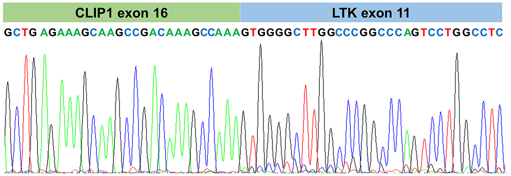
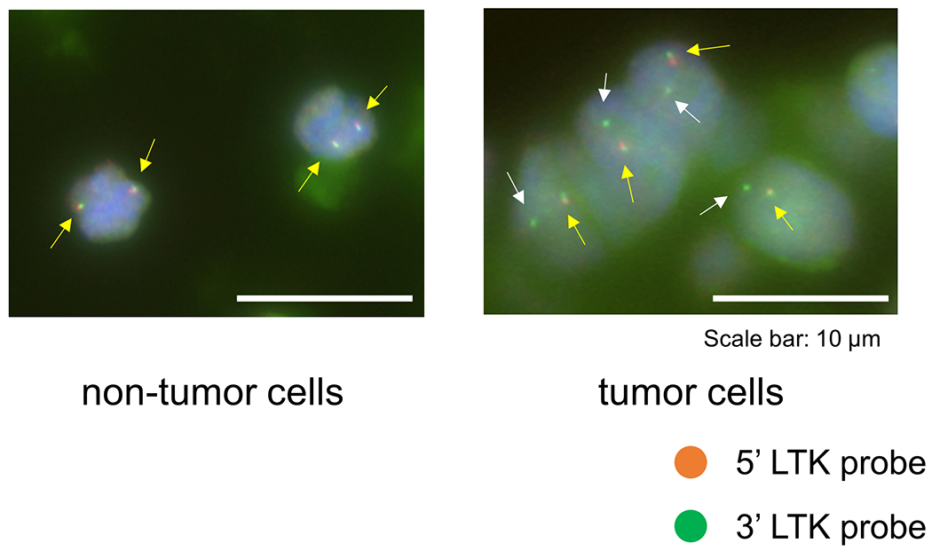
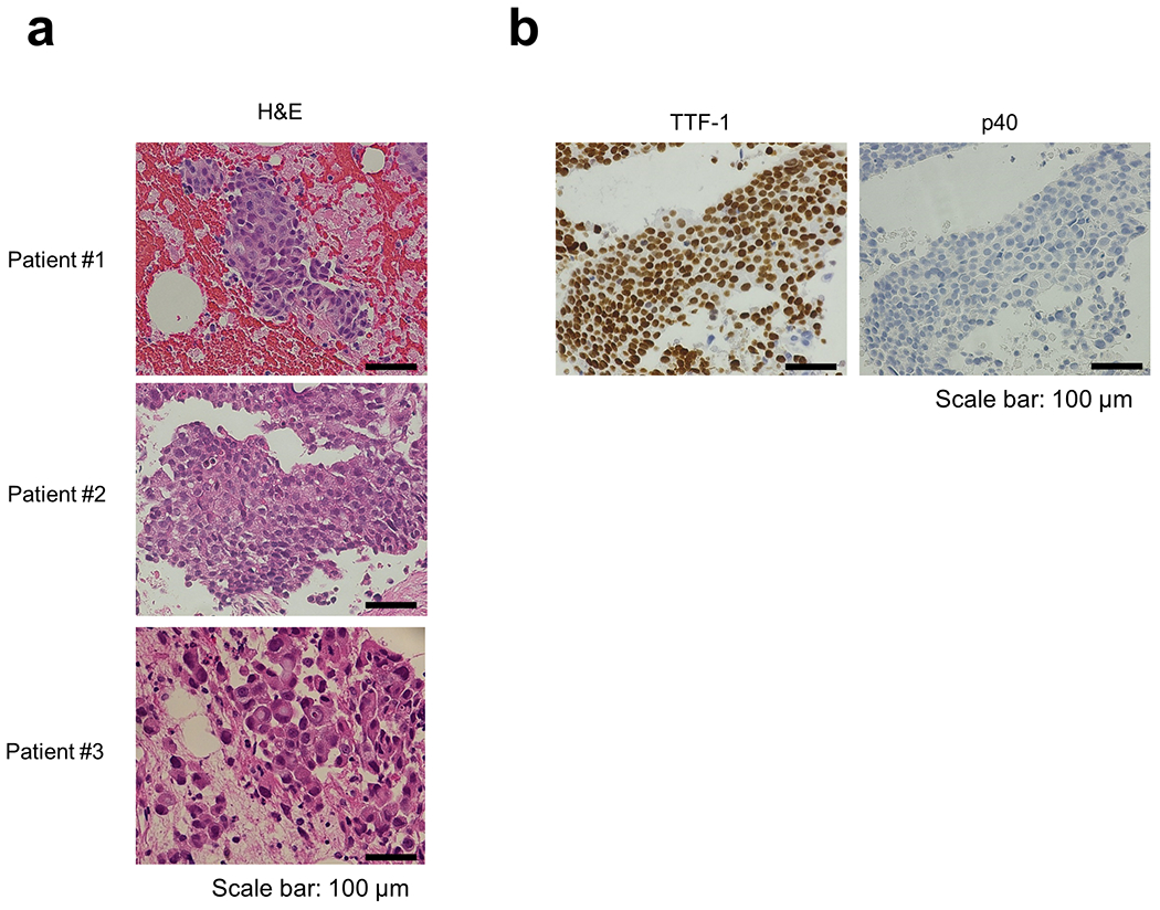
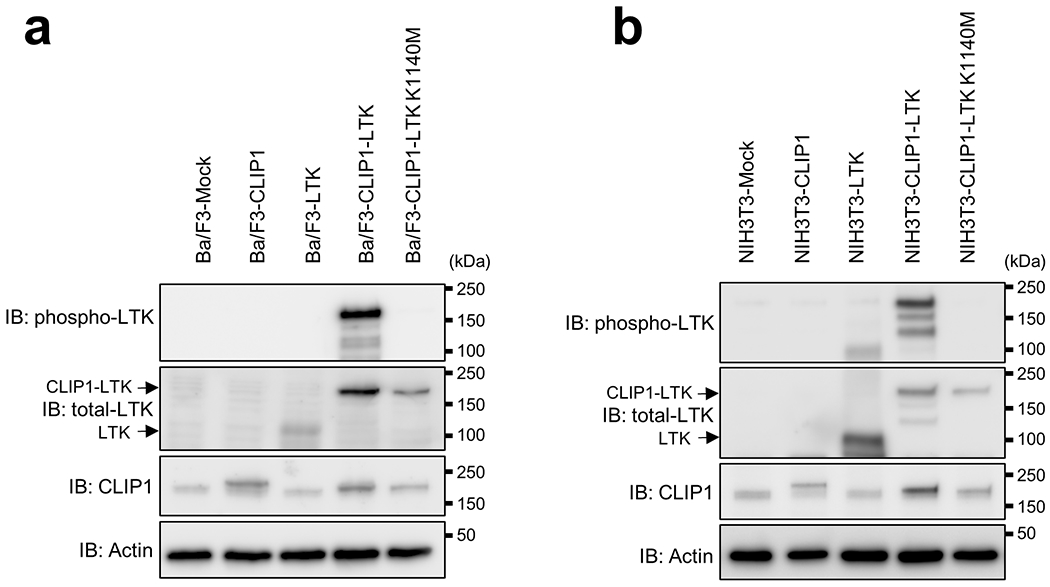
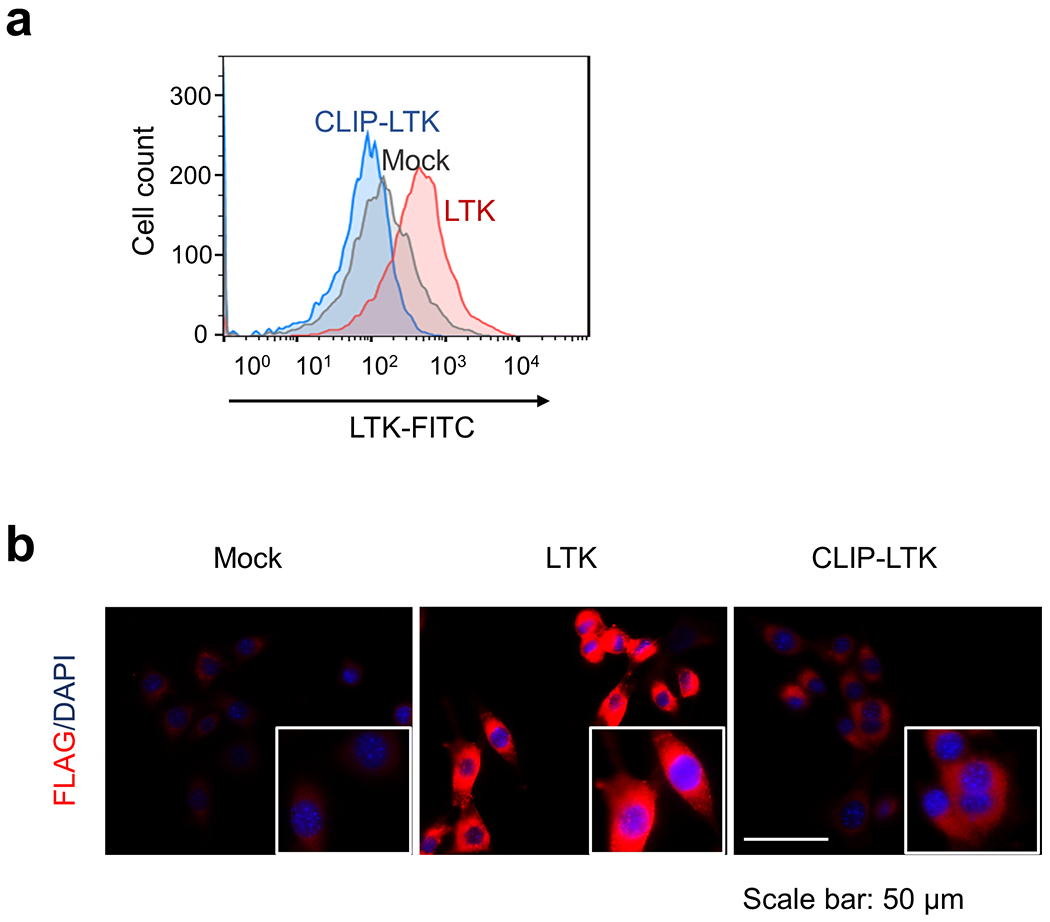
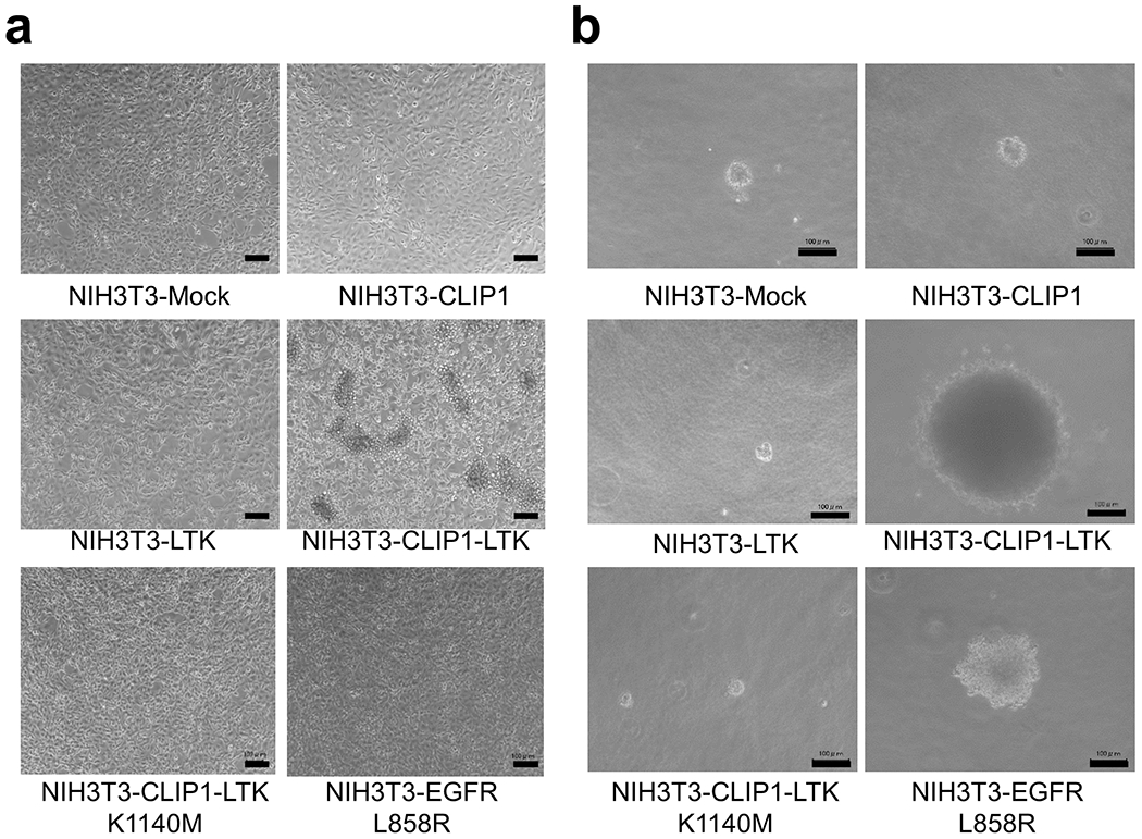
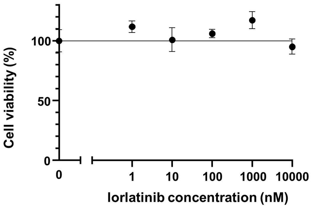
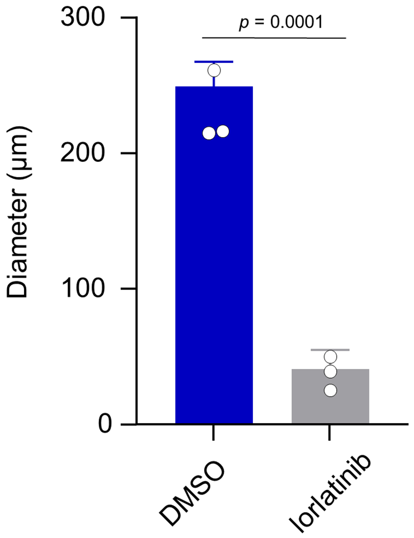
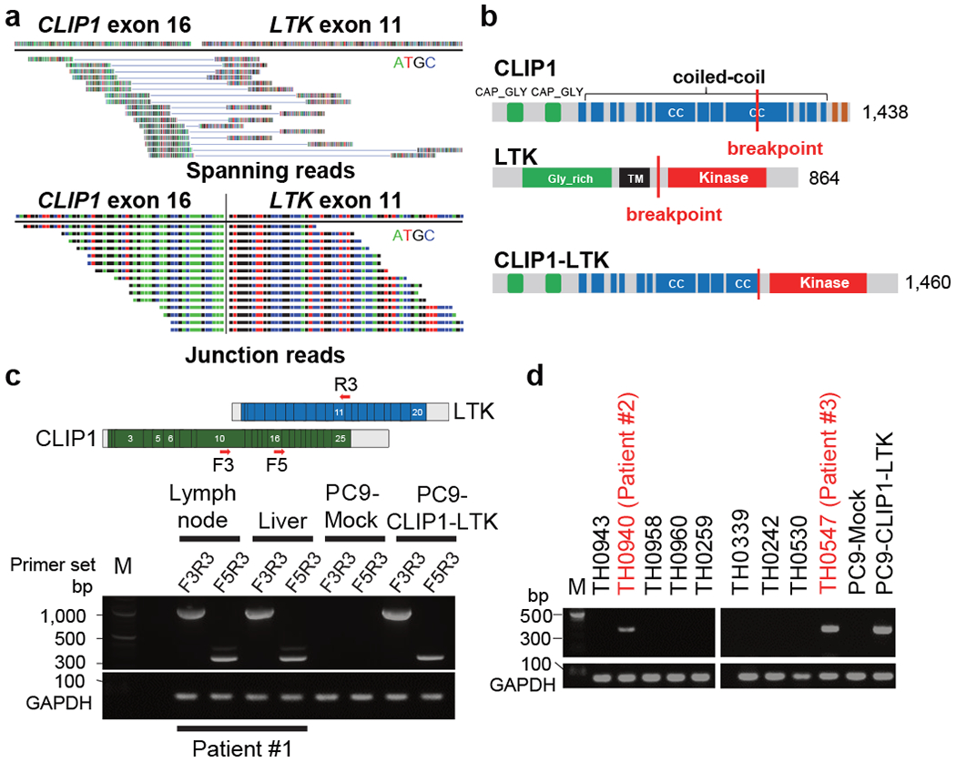
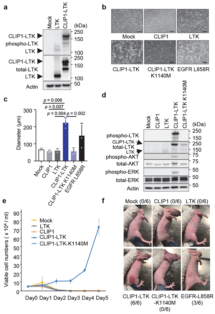
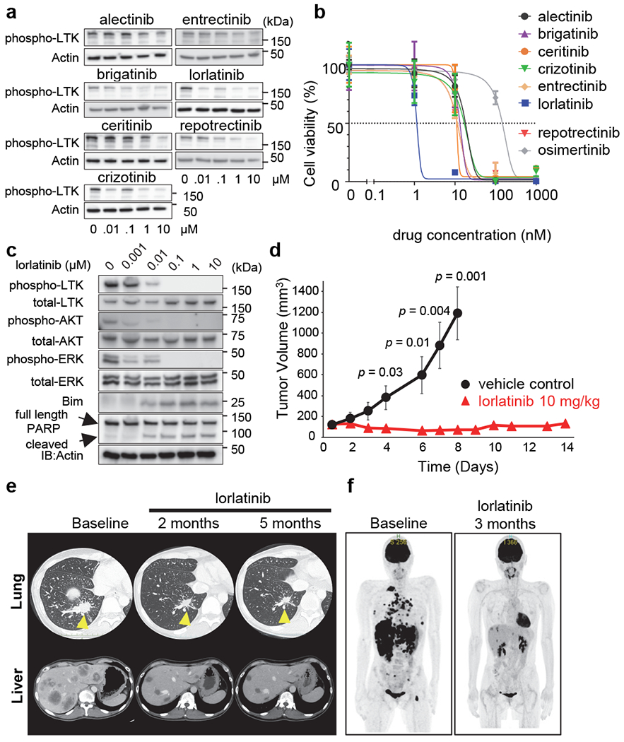
Lung cancer is one of the most aggressive tumour types. Targeted therapies stratified by oncogenic drivers have substantially improved therapeutic outcomes in patients with non-small-cell lung cancer (NSCLC)1. However, such oncogenic drivers are not found in 25-40% of cases of lung adenocarcinoma, the most common histological subtype of NSCLC2. Here we identify a novel fusion transcript of CLIP1 and LTK using whole-transcriptome sequencing in a multi-institutional genome screening platform (LC-SCRUM-Asia, UMIN000036871). The CLIP1-LTK fusion was present in 0.4% of NSCLCs and was mutually exclusive with other known oncogenic drivers. We show that kinase activity of the CLIP1-LTK fusion protein is constitutively activated and has transformation potential. Treatment of Ba/F3 cells expressing CLIP1-LTK with lorlatinib, an ALK inhibitor, inhibited CLIP1-LTK kinase activity, suppressed proliferation and induced apoptosis. One patient with NSCLC harbouring the CLIP1-LTK fusion showed a good clinical response to lorlatinib treatment. To our knowledge, this is the first description of LTK alterations with oncogenic activity in cancers. These results identify the CLIP1-LTK fusion as a target in NSCLC that could be treated with lorlatinib.


Figures

Extended Data Fig. 1:
Extended Data Fig. 1:. Electropherogram showing Sanger sequencing of the CLIP1-LTK fusion transcript.
cDNAs were generated from RNAs isolated from patient tumors and amplified by RT-PCR with CLIP1-LTK F3 and R3 primers. PCR products were directly sequenced using respective primers.
Extended Data Fig. 2:
Extended Data Fig. 2:. LTK break-apart FISH assay.
Non-tumor cells from Patient #1 showed orange (5’)-green (3’) fused signals (yellow arrows), while 82% (41/50) of scored tumor cells showed one fused (yellow arrows) and at least one green (3’) signals (white arrows), which indicates the presence of LTK rearrangement. Bars = 10 μm.
Extended Data Fig. 3:
Extended Data Fig. 3:. Histological findings of tumors positive for the CLIP1-LTK fusion.
a, Hematoxylin and eosin (H&E) stained images. Samples from Patient #1 and #3 were diagnosed with adenocarcinoma morphologically. Samples from Patient #2 was diagnosed with NSCLC. Bars = 100 μm. b, Immunohistochemical analysis of samples from Patient #2. TTF-1 positive (upper panel) and p40 negative (lower panel) staining supported the diagnosis of NSCLC favor adenocarcinoma. Bars = 100 μm.
Extended Data Fig. 4:
Extended Data Fig. 4:. Analysis of Ba/F3 (a) and NIH3T3 (b) cells stably transduced using MIGR1 IRES-GFP vectors harboring indicated constructs.
GFP-positive cells were sorted and expanded and then cell extracts were immunoblotted with antibodies indicated at the left side of each graph.
Extended Data Fig. 5:
Extended Data Fig. 5:. Cellular localization of LTK and CLIP1-LTK.
a, NIH3T3-Mock NIH3T3-LTK, or NIH3T3-CLIP1-LTK cells were stained with primary antibody specific for LTK, conjugated with Alexa Fluor 488, and subjected to flow cytometry analysis. Viable cells were gated as shown in Supplementary Information Figure 2. b, NIH3T3-Mock NIH3T3-LTK, or NIH3T3-CLIP1-LTK cells were fixed, permeabilized, and stained with anti-DDDDK-tag antibody conjugated to Alexa Fluor 594. Cells were subjected to immunofluorescence analysis.
Extended Data Fig. 6:
Extended Data Fig. 6:. Light microscopy images of indicated stably-transduced NIH3T3 cells.
a, Cells were plated in 10 cm plates at 2 x 10⁵ cells/ml and cultured in DMEM supplemented with 10% FBS and P/S for 2-3 days until cells reached 100% confluency. Bars = 100 μm. b, Cells were plated in 6-well plates at 1 x 10⁴ cells/ml and cultured in a soft agar medium for 14 days. Bars = 100 μm.
Extended Data Fig. 7:
Extended Data Fig. 7:. Analysis of viability of Ba/F3-CLIP1-LTK cells.
Ba/F3-CLIP1-LTK cells were treated with various concentrations of lorlatinib in the presence of 5% WEHI medium as a source of IL-3. Results are shown as an average ± standard deviation from three independent experiments.
Extended Data Fig. 8:
Extended Data Fig. 8:. Lorlatinib suppresses anchorage-independent growth of NIH3T3-CLIP1-LTK cell colonies.
Colony diameters were measured in lorlatinib- versus vehicle (DMSO)-treated cells and shown as average ± standard deviation from three independent experiments.
Fig. 1
Fig. 1. Identification of the CLIP1-LTK fusion.
a, Spanning (top) and junction (bottom) reads for the CLIP1-LTK fusion transcript, as detected by WTS. b, Schematic representations of WT CLIP1 (top), WT LTK (middle), and the CLIP1-LTK fusion protein (bottom). CC, coiled-coil; TM, transmembrane. c, Detection by RT-PCR of CLIP1-LTK transcripts in two metastatic sites, the supraclavicular lymph node and liver from Patient #1. Primer locations are indicated at the top. cDNA isolated from PC9 cells expressing the CLIP1-LTK fusion or Mock cells (see Methods) served as respective positive and negative controls. GAPDH transcripts serve as an internal control. M, 100-bp DNA ladder marker. d, Screening of lung cancer specimens for the CLIP1-LTK transcript using the F5 and R3 primer set. Shown are representative panels of RT-PCR screening that contain two positive samples (#2 and #3), as well as GAPDH.
Fig. 2
Fig. 2. Transforming activity of the CLIP1-LTK fusion.
a, Constitutive tyrosine kinase activity of CLIP1-LTK fusion protein. NIH3T3 cells were transiently transfected with empty vector or WT LTK or CLIP1-LTK expression plasmids, and 48 hours later cell extracts were immunoblotted with anti-phospho LTK antibodies. b, Light microscopy images of indicated stably-transduced NIH3T3 cells. Cells were plated in 10 cm plates at 2 x 10⁵ cells/ml and cultured for 1-2 days until cells reached 50-60 % confluency. Bars = 100 μm. c, A soft agar colony formation assay of indicated NIH3T3 stably-transduced cells. Colony diameters were measured and average values ± standard deviations from three independent experiments are shown. d, Activation of AKT and ERK downstream of CLIP1-LTK. Ba/F3 cells transduced with either Mock, CLIP1, LTK, CLIP1-LTK, or CLIP1-LTK-K1140M were cultured at 5 x 10⁵ cells/ml in the absence of 5% WEHI medium for 12 hours. Cell extracts were then subjected to western blotting analysis with indicated antibodies. e, IL-3-independent growth of Ba/F3-CLIP1-LTK cells. Prior to plating, Ba/F3 cells transduced with indicated constructs and growing in 5% WEHI medium were washed and resuspended in RPMI medium at 5 x 10⁴ cells/ml. Living cells were counted daily and shown as an average ± standard deviation from three independent experiments. f, CLIP1-LTK has tumor transforming activity. NIH3T3 cells transduced with transduced with either Mock, CLIP1, LTK, CLIP1-LTK, CLIP1-LTK-K1140M, or EGFR-L858R were injected into the flanks of nude mice and observed for tumor growth. Representative pictures taken at day 10 following injection are shown. The numbers of tumors induced in the injected animals are shown in parentheses.
Fig. 3
Fig. 3. Lorlatinib inhibits CLIP1-LTK kinase activity and suppresses tumor growth.
a, Screening for CLIP1-LTK inhibitors. NIH3T3 cells were transiently transfected with pCMV3-CLIP1-LTK, cultured 24 hours, and then treated 6 hours with indicated TKIs. Cell extracts were subjected to western blotting analysis (using antibodies against phospho-LTK and Actin). b, Viability assays of inhibitor-treated cells. Ba/F3-CLIP-LTK cells were treated 48 hours with increasing concentrations of indicated chemicals and viability was measured using the Cell Counting Kit-8. Results are shown as an average ± standard deviation from three independent experiments. c, Inhibition of CLIP-LTK kinase activity and downstream signaling by lorlatinib. Ba/F3-CLIP-LTK cells (4 x 10⁵ cells/well in 6 well plates) were treated 24 hours with increasing lorlatinib concentrations. Cell extracts were then subjected to western blotting analysis with indicated antibodies. d, Inhibition of tumor growth by lorlatinib in vivo. NIH3T3-CLIP-LTK cells (3 x 10⁶ cells) were transplanted into the flanks of nude mice. When the mean tumor volume reached 100-200 mm3, mice were randomized into two groups and treated with either lorlatinib (10 mg/kg once daily) or vehicle control by oral gavage for 14 days and 8 days, respectively. Tumor volumes are shown as an average ± standard error (n=6 for each group). e, Time course CT scan images of right lung (upper panels) and liver (lower panels) in Patient #1, who had been treated with lorlatinib orally at 100 mg daily. f, Time course PET scan images of the whole body.

Comment in

References

    1. Konig D, Savic Prince S & Rothschild SI Targeted Therapy in Advanced and Metastatic Non-Small Cell Lung Cancer. An Update on Treatment of the Most Important Actionable Oncogenic Driver Alterations. Cancers (Basel) 13, doi:10.3390/cancers13040804 (2021). - DOI - PMC - PubMed
    1. Saito M et al. Gene aberrations for precision medicine against lung adenocarcinoma. Cancer Sci 107, 713–720, doi:10.1111/cas.12941 (2016). - DOI - PMC - PubMed
    1. Cancer Genome Atlas Research, N. Comprehensive molecular profiling of lung adenocarcinoma. Nature 511, 543–550, doi:10.1038/nature13385 (2014). - DOI - PMC - PubMed
    1. Fernandez-Cuesta L et al. CD74-NRG1 fusions in lung adenocarcinoma. Cancer Discov 4, 415–422, doi:10.1158/2159-8290.CD-13-0633 (2014). - DOI - PubMed
    1. National Comprehensive Cancer Network. Non-Small Cell Lung Cancer (Version 4.2021), <https://www.nccn.org/professionals/physician_gls/pdf/nscl.pdf> (2021).

Publication types

MeSH terms