. 2021 Nov 8:12:752168.
doi: 10.3389/fimmu.2021.752168. eCollection 2021.

Multi-Antigen Outer Membrane Vesicle Engineering to Develop Polyvalent Vaccines: The Staphylococcus aureus Case

Affiliations

Multi-Antigen Outer Membrane Vesicle Engineering to Develop Polyvalent Vaccines: The Staphylococcus aureus Case

Enrico König et al. Front Immunol. .

Abstract

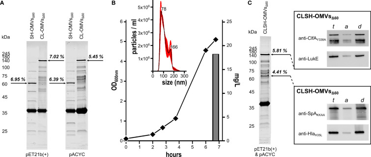
Modification of surface antigens and differential expression of virulence factors are frequent strategies pathogens adopt to escape the host immune system. These escape mechanisms make pathogens a "moving target" for our immune system and represent a challenge for the development of vaccines, which require more than one antigen to be efficacious. Therefore, the availability of strategies, which simplify vaccine design, is highly desirable. Bacterial Outer Membrane Vesicles (OMVs) are a promising vaccine platform for their built-in adjuvanticity, ease of purification and flexibility to be engineered with foreign proteins. However, data on if and how OMVs can be engineered with multiple antigens is limited. In this work, we report a multi-antigen expression strategy based on the co-expression of two chimeras, each constituted by head-to-tail fusions of immunogenic proteins, in the same OMV-producing strain. We tested the strategy to develop a vaccine against Staphylococcus aureus, a Gram-positive human pathogen responsible for a large number of community and hospital-acquired diseases. Here we describe an OMV-based vaccine in which four S. aureus virulent factors, ClfAY338A, LukE, SpAKKAA and HlaH35L have been co-expressed in the same OMVs (CLSH-OMVsΔ60). The vaccine elicited antigen-specific antibodies with functional activity, as judged by their capacity to promote opsonophagocytosis and to inhibit Hla-mediated hemolysis, LukED-mediated leukocyte killing, and ClfA-mediated S. aureus binding to fibrinogen. Mice vaccinated with CLSH-OMVsΔ60 were robustly protected from S. aureus challenge in the skin, sepsis and kidney abscess models. This study not only describes a generalized approach to develop easy-to-produce and inexpensive multi-component vaccines, but also proposes a new tetravalent vaccine candidate ready to move to development.

Keywords: OMV engineering; Staphylococcus aureus; chimeric proteins; multivalent vaccines; outer membrane vesicles (OMVs).


Conflict of interest statement

Author FB was employed by company GlaxoSmithKline Vaccines. GG, EK, IZ, CI, and LFa are coinventors of a patent on OMVs. AGr and GG are involved in a biotech company interested in exploiting the OMV platform. The remaining authors declare that the research was conducted in the absence of any commercial or financial relationships that could be construed as a potential conflict of interest.

Figures

Figure 1
Figure 1
Expression of Staphylococcal antigen fusions in OMVs from E. coli BL21(DE3)Δ60. (A) OMVs from E. coli BL21(DE3)Δ60 strains expressing heterologous antigen fusions SpAKKAA-HlaH35L (SH-OMVsΔ60) and ClfAY338A-LukE (CL-OMVsΔ60) as lipoproteins using either pET21b(+) or pACYC as plasmid backbone were purified from culture supernatants as described in Materials and Methods. Aliquots (10 μg of total OMV proteins) were analyzed by SDS-PAGE. (B) Growth curve, OMV production yield and particle size analysis of CLSH-OMVsΔ60 grown in a 3-L bioreactor (see Materials and Methods for details). The size of CLSH-OMVsΔ60 was determined using a NanoSight NS300 (Malvern Panalytical). The graph shows the average of three independent measurements and standard error as calculated by the manufacturer’s software. Numbers indicate the particle size of main peaks. (C) OMVs from E. coli BL21(DE3)Δ60 co-expressing the two heterologous antigen fusions ClfAY338A-LukE and SpAKKAA-HlaH35L as lipoproteins (CLSH-OMVsΔ60) and analysis of protein lipidation by Triton X-114 extraction of OMV proteins. ClfAY338A-LukE and SpAKKAA-HlaH35L were co-expressed using pET21b(+) and pACYC, respectively, and purified and analyzed by SDS-PAGE as stated for panel (A) For analysis of protein lipidation, CLSH-OMVsΔ60 expressing heterologous protein fusions in the membrane were dissolved by adding 1% Triton X-114 at 4°C and subsequently aqueous and detergent phases were partitioned by centrifugation. Unfractionated total proteins from CLSH-OMVsΔ60 (t), hydrophilic proteins in the aqueous phase (a), and hydrophobic proteins in the detergent phase (d) were precipitated with chloroform-methanol and separated by SDS-PAGE. Finally, proteins were transferred onto nitrocellulose filters and the presence of the heterologous antigen fusions in either the aqueous or detergent phase was detected by Western blot using antigen-specific antibodies. The numbers next to each arrow (A, C) represent the percentage of each recombinant chimera over total OMV protein content as estimated by densitometry using Image Studio Lite Ver 5.2.
Figure 2
Figure 2
Immunogenicity of CLSH-OMVsΔ60. (A) Antigen-specific antibody titers. Groups of 8 female CD1 mice were immunized i.p. (left panel) or i.m. (right panel) 3 times at 2-week intervals with 20 μg of CLSH-OMVsΔ60 formulated with or without Alum (filled and open symbols, respectively). Sera were collected 7 days after the last immunization and IgG titers were analyzed by ELISA using plates coated with the respective purified recombinant protein (300 ng/well). Each data point represents the antibody titer from a single mouse. Mean ± s.e.m. is shown. (B) Opsonophagocytosis killing (OPK). 500 CFUs of S. aureus Newman strain grown in TSB at 37°C were incubated with serially diluted immune sera for 45 minutes at RT. Then, 3×10⁵ HL-60 cells differentiated into neutrophils and guinea pig complement were added to each reaction for 90 minutes at 37°C (T90). Bacterial killing refers to the CFUs of the negative control (bacteria + HL-60 cells + complement without serum) set as 0% killing and was calculated as follows: [CFUs of the negative control at T90 - CFUs of each sample at T90]/CFUs of the negative control at T90]. Data are reported as mean ± s. d. of two independent experiments. HC: heat-inactivated complement. (C) Inhibition of fibrinogen binding. S. aureus Newman strain (1×10⁷ CFUs/well) was pre-incubated for 30 minutes at RT with serially diluted pooled sera from mice immunized with either 20 μg of “empty” OMVsΔ60 (grey bars) or 20 μg of CLSH-OMVsΔ60 (black bars). Mixtures were transferred to a 96-well plate previously coated with human fibrinogen (10 µg/ml) and incubated for 1 hour at 37°C. Adherent bacteria were fixed with formaldehyde and then stained with crystal violet. Adherence to fibrinogen was calculated as a percentage of values measured in control wells lacking serum (= 100%), and inhibition of binding to fibrinogen was calculated by subtracting the adherence percentage values from the control (= 100% - x %). The graph shows the mean ± s. d. of three independent experiments. (D) Inhibition of leukocidin-mediated cytotoxicity. HL-60 cells differentiated into neutrophils (5×10⁵/well) were incubated for 24 hours at 37°C with LukED (200 nM) in presence of serially two-fold diluted sera from mice immunized with 20 μg of CLSH-OMVsΔ60 (black bars), “empty” OMVsΔ60 (grey bars) or with Alum (white bars). For assessment of cell viability, the XTT Assay reagent was added and after 16-hour incubation at 37°C the absorbance was read at 470 nm. Data are reported as mean ± s. d. of two independent experiments. (E) Inhibition of Hla-mediated hemolysis. Serially two-fold diluted sera from mice immunized with either 20 μg of “empty” OMVsΔ60 (grey bars) or 20 μg of CLSH-OMVsΔ60 (black bars) were pre-incubated with recombinant Hla for 20 minutes at RT and then with rabbit erythrocytes for 30 minutes at 37°C. Hla hemolytic activity was calculated as percentage of hemolytic activity obtained for rabbit erythrocytes incubated with water (100% hemolysis). The graph shows the mean ± s. d. of three independent experiments.
Figure 3
Figure 3
In vivo protective activity of OMVsΔ60 expressing S. aureus antigens. (A) Sepsis model. Groups of 16 female CD1 mice were immunized three times at 2-week intervals with Alum alone (circles), “empty” OMVsΔ60 (triangles) or CLSH-OMVsΔ60 (diamonds), formulated in Alum. After 2 weeks, mice were infected i.p. with 3×10⁸ CFUs of S. aureus Newman strain. Animal health was monitored over a period of 7 days, assigning a “pain score” from 1 to 4. Animals which reached a pain score = 4 were sacrificed. The three graphs show the pain score (left), survival at day 7 (center) and the survival over time (Kaplan-Meier curve) (right). See text for definition of “survival”. Statistical analysis for the pain score plot was performed using Mann-Whitney test. Median is shown. **P = 0.0015. Statistical analysis for the Kaplan–Meier plot was performed using the log rank test. *P = 0.0253; ***P = 0.0001. (B) Skin model. Groups of 16 CD1 female mice were immunized three times at 2-week intervals with Alum alone (circles), “empty” OMVsΔ60 (triangles) and CLSH-OMVsΔ60 (diamonds). At day 14 after the third immunization, mice were s.c. infected with 5×10⁷ CFUs of S. aureus Newman strain. Abscess size was monitored once a day for 14 days. Mean ± s. d. is shown. Skin abscess areas of vaccinated and control animals were tested for significance by means of repeated ANOVA measures using the R base function aov(). (C) Renal abscess model. Groups of 16 CD1 female mice were immunized three times at 2-week intervals with Alum alone (circles), “empty” OMVsΔ60 (triangles) and CLSH-OMVsΔ60 (diamonds). Ten days after the last immunization, mice were infected i.v. with a sub-lethal dose of S. aureus Newman strain (1×10⁷ CFUs) and 4 days afterward, mice were sacrificed, kidneys collected and homogenized in PBS, and aliquots were plated on agar media for CFUs determination. Statistical analysis was performed using two-tailed Student’s t-test. Geometric mean ± 95% confidence interval is shown. *P < 0.05.
Figure 4
Figure 4
Expression of chimeras in OMVsΔ60. (A) E. coli BL21(DE3)Δ60 was transformed with plasmids expressing the following chimeras: FhuD2-HlaH35L, FhuD2-LukE, LukE-HlaH35L, SpAKKAA-HlaH35L and ClfAY338A-LukE. One recombinant clone from each transformation was inoculated in liquid culture and the corresponding OMVs, named FH-OMVsΔ60, FL-OMVsΔ60, LH-OMVsΔ60, SH-OMVsΔ60 and CL-OMVsΔ60, respectively, were purified from each culture supernatants. Purified OMVs were analyzed by SDS-PAGE (10 μg total OMV proteins) (left panel). The right panel reports the SDS-PAGE analysis of OMVs from E. coli BL21(DE3)Δ60 co-transformed with plasmids expressing either ClfAY338A-LukE and FhuD2-HlaH35L (CLFH-OMVsΔ60) or FhuD2-LukE and SpAKKAA-HlaH35L (FLSH-OMVsΔ60) using pET21b(+) and pACYC, respectively (see Materials and Methods for details). (B) A group of five mice were immunized three times two weeks apart with CLFH-OMVsΔ60. After two weeks from the last immunization sera were collected and pooled and the ELISA titers were measured coating the plates with recombinant proteins.

References

    1. Plotkin SA, Orenstein WA, Offit PA, Edwards KM. eds. Vaccines. 7th ed. Philadelphia: Elsevier; (2018).
    1. McCarthy PC, Sharyan A, Moghaddam LS. Meningococcal Vaccines: Current Status and Emerging Strategies. Vaccines (2018) 6:12. doi: 10.3390/vaccines6010012 - DOI - PMC - PubMed
    1. Ellis TN, Kuehn MJ. Virulence and Immunomodulatory Roles of Bacterial Outer Membrane Vesicles. Microbiol Mol Biol Rev (2010) 74:81–94. doi: 10.1128/MMBR.00031-09 - DOI - PMC - PubMed
    1. Klimentovà J, Stulík J. Methods of Isolation and Purification of Outer Membrane Vesicles From Gram-Negative Bacteria. Microbiol Res (2015) 170:1–9. doi: 10.1016/j.micres.2014.09.006 - DOI - PubMed
    1. Kesty NC, Kuehn MJ. Incorporation of Heterologous Outer Membrane and Periplasmic Proteins Into Escherichia Coli Outer Membrane Vesicles. J Biol Chem (2004) 279:2069–76. doi: 10.1074/jbc.M307628200 - DOI - PMC - PubMed

Publication types