

SLC41A3 Exhibits as a Carcinoma Biomarker and Promoter in Liver Hepatocellular Carcinoma
- PMID: 34819993
- PMCID: PMC8608493
- DOI: 10.1155/2021/8556888
SLC41A3 Exhibits as a Carcinoma Biomarker and Promoter in Liver Hepatocellular Carcinoma
Abstract
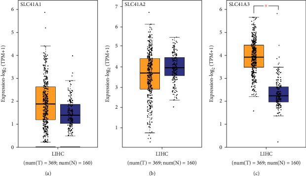
Liver Hepatocellular Carcinoma (LIHC) is the fifth widely occurred carcinoma, which is thought to be the second primary contributor of carcinoma-associated death. There are almost 788,000 death tolls worldwide. Solute carrier family 41 member 3 (SLC41A3) is a member of solute carrier family 41, and it is the key point of numerous researches. Our research attempted to explore the links between SLC41A3 and LIHC through public databases. Higher expression of SLC41A3 displayed an intimate association with higher pathological stages and poorer prognosis. GO and KEGG analysis revealed the possible regulatory pathways of SLC41A3. Additionally, we carried out cell functional experiments to determine the expression of SLC41A3 in the cell lines of LIHC, as well as the effects of its silence on cell proliferation, migration, and invasion. Our data showed that SLC41A3 was greatly increased in the cell lines of LIHC. Moreover, silencing SLC41A3 impeded LIHC cell proliferation, migration, and invasion in vitro. Collectively, our study demonstrated that highly expressed SLC41A3 was a probable indication of LIHC occurrence, and SLC41A3 could be regarded as a prospective target in the treatment of LIHC.
Copyright © 2021 Qimeng Chang et al.
Conflict of interest statement
The authors declare that they have no conflicts of interest.
Figures


Similar articles
-
Construction of liver hepatocellular carcinoma-specific lncRNA-miRNA-mRNA network based on bioinformatics analysis.PLoS One. 2021 Apr 16;16(4):e0249881. doi: 10.1371/journal.pone.0249881. eCollection 2021. PLoS One. 2021. PMID: 33861762 Free PMC article.
-
A Pyroptosis-Related Gene Signature to Predict Patients' Prognosis and Immune Landscape in Liver Hepatocellular Carcinoma.Comput Math Methods Med. 2022 Feb 16;2022:1258480. doi: 10.1155/2022/1258480. eCollection 2022. Comput Math Methods Med. 2022. PMID: 35242200 Free PMC article.
-
Upregulation Mitochondrial Carrier 1 (MTCH1) Is Associated with Cell Proliferation, Invasion, and Migration of Liver Hepatocellular Carcinoma.Biomed Res Int. 2021 Jun 7;2021:9911784. doi: 10.1155/2021/9911784. eCollection 2021. Biomed Res Int. 2021. PMID: 34195286 Free PMC article.
-
Diagnostic significance and carcinogenic mechanism of pan-cancer gene POU5F1 in liver hepatocellular carcinoma.Cancer Med. 2020 Dec;9(23):8782-8800. doi: 10.1002/cam4.3486. Epub 2020 Sep 26. Cancer Med. 2020. PMID: 32978904 Free PMC article.
-
Long Non-Coding RNAs as Mediators of Tumor Microenvironment and Liver Cancer Cell Communication.Int J Mol Sci. 2018 Nov 24;19(12):3742. doi: 10.3390/ijms19123742. Int J Mol Sci. 2018. PMID: 30477236 Free PMC article. Review.
Cited by
-
Plasma Exosome Gene Signature Differentiates Colon Cancer from Healthy Controls.Ann Surg Oncol. 2023 Jun;30(6):3833-3844. doi: 10.1245/s10434-023-13219-7. Epub 2023 Mar 2. Ann Surg Oncol. 2023. PMID: 36864326 Free PMC article.
-
SLC41A1 overexpression correlates with immune cell infiltration in HCC and promotes its malignant progression.Int J Med Sci. 2024 Nov 11;21(15):3069-3082. doi: 10.7150/ijms.100155. eCollection 2024. Int J Med Sci. 2024. PMID: 39628683 Free PMC article.
-
Optimizing Prognostic Predictions in Liver Cancer with Machine Learning and Survival Analysis.Entropy (Basel). 2024 Sep 7;26(9):767. doi: 10.3390/e26090767. Entropy (Basel). 2024. PMID: 39330100 Free PMC article.
-
Genotypic and phenotypic consequences of domestication in dogs.bioRxiv [Preprint]. 2024 Nov 30:2024.05.01.592072. doi: 10.1101/2024.05.01.592072. bioRxiv. 2024. PMID: 38746159 Free PMC article. Preprint.
References
MeSH terms
Substances
LinkOut - more resources
Full Text Sources
Medical
Molecular Biology Databases
