. 2022 Jan;23(1):12.
doi: 10.3892/ol.2021.13130. Epub 2021 Nov 11.

Diagnostic and prognostic value of serum miR-145 and vascular endothelial growth factor in non-small cell lung cancer

Affiliations

Diagnostic and prognostic value of serum miR-145 and vascular endothelial growth factor in non-small cell lung cancer

Sarayut Lucien Geater et al. Oncol Lett. 2022 Jan.

Abstract

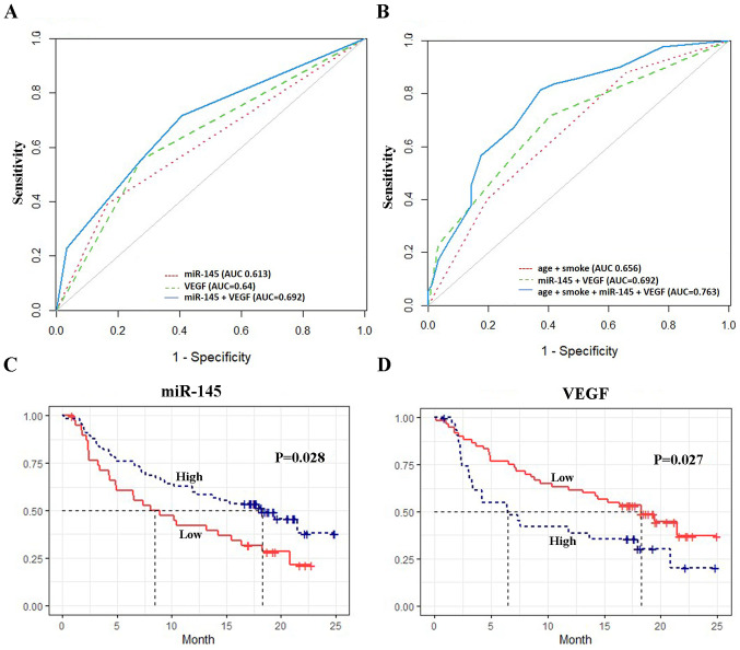
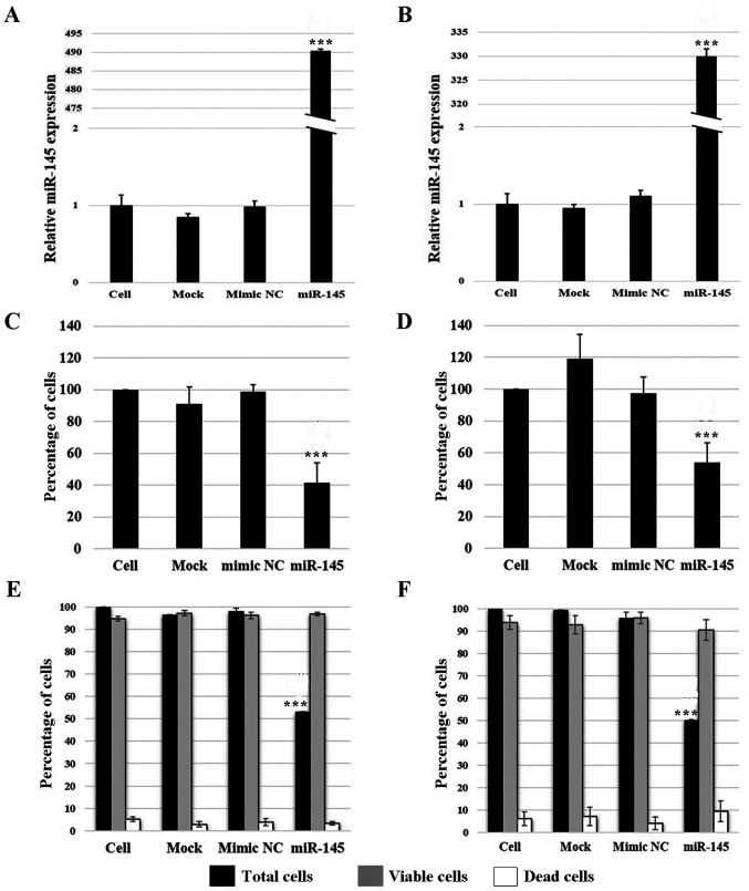
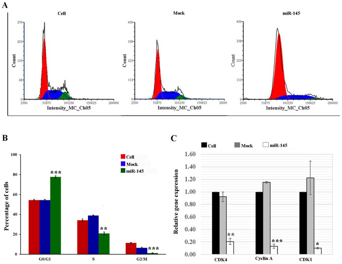
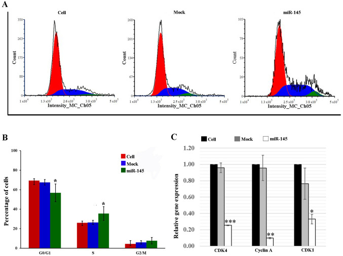
Previous studies have reported the diagnostic and prognostic value of serum microRNA (miR)-145 and vascular endothelial growth factor (VEGF) levels in various types of cancer; however, their clinical use in non-small cell lung cancer (NSCLC) remains unclear. The present study included 215 patients, 106 with NSCLC and 109 with other lung diseases (OLDs). miR-145 expression levels were determined using reverse transcription-quantitative PCR (RT-qPCR) and VEGF levels were measured using an ELISA. The diagnostic performance was assessed using a receiver operating characteristic curve and area under the curve (AUC) analysis. A Kaplan-Meier survival curve and Cox regression analysis were employed to evaluate the prognostic significance of the markers. The biological function of miR-145 was examined in A549 and H1792 cell lines. The effects of miR-145 on cell proliferation of NSCLC cells were evaluated by flow cytometry, and the expression levels of miR-145 and cell cycle-related genes were determined by RT-qPCR. The results revealed that miR-145 and VEGF exhibited fair diagnostic performance [AUC, 0.61 (95% CI, 0.55-0.68) and AUC, 0.64 (95% CI, 0.57-0.71), respectively]. Combining age and smoking status with miR-145 and VEGF provided the best model for differentiating patients with NSCLC from those with OLDs (AUC, 0.76; 95% CI, 0.69-0.83). Furthermore, low serum miR-145 levels were associated with poor overall survival [hazard ratio (HR), 0.48; 95% CI, 0.27-0.85], whereas high VEGF levels were not associated with poor overall survival (HR, 1.47; 95% CI, 0.81-2.68). In addition, the results of the in vitro experiments indicated that miR-145 decreased cell proliferation via the induction of cell cycle arrest. In conclusion, the findings of the present study suggested that combining miR-145 and VEGF levels with clinical risk factors may be a potential diagnostic scheme for NSCLC. In addition, serum miR-145 may be used as a prognostic marker. These results indicated that miR-145 may function as a tumor suppressor in NSCLC.

Keywords: NSCLC; VEGF; diagnostic; miR-145; prognostic; serum.


Conflict of interest statement

The authors declare that they have no competing interests.

Figures

Figure 1.
Figure 1.
(A and B) Receiver operating characteristic curve analysis for the diagnosis of NSCLC. Kaplan-Meier curves of overall survival of patients with NSCLC according to (C) miR-145 expression and (D) VEGF levels. AUC, area under the curve; NSCLC, non-small cell lung cancer; miR, microRNA; VEGF, vascular endothelial growth factor.
Figure 2.
Figure 2.
Reverse transcription-quantitative PCR analysis was performed to detect miR-145 expression in (A) A549 and (B) H1792 cells. Effect of upregulated miR-145 expression on viability was determined using a trypan blue assay in (C) A549 and (D) H1792 cells, and using a viability assay in (E) A549 and (F) H1792 cells. The cell group was set as 100%. All experiments were performed in triplicate on cell-culture passages 10-30. Data are presented as the mean ± SD of three independent experiments. OL-23-01-13130P≤0.001 vs. cell group. miR, microRNA; NC, negative control.
Figure 3.
Figure 3.
(A and B) Cell cycle progression was analyzed via flow cytometry in A549 cells. (C) Expression of cell cycle regulatory genes was determined using reverse transcription-quantitative PCR. All experiments were performed in triplicate on cell-culture passages 10–30. Data are presented as the mean ± SD from three independent experiments. ***P≤0.001, **P≤0.01, *P≤0.05 vs. cell group. miR, microRNA; NC, negative control.
Figure 4.
Figure 4.
(A and B) Cell cycle progression was determined using flow cytometry in H1792 cells. (C) Expression of cell cycle regulatory genes was determined using reverse transcription-quantitative PCR. All experiments were performed in triplicate on cell-culture passages 10–30. Data are presented as the mean ± SD from three independent experiments. ***P≤0.001, **P≤0.01, *P≤0.05 vs. cell group. miR, microRNA; NC, negative control.

Similar articles

Cited by

References

    1. Sung H, Ferlay J, Siegel RL, Laversanne M, Soerjomataram I, Jemal A, Bray F. Global cancer statistics 2020: GLOBOCAN estimates of incidence and mortality worldwide for 36 cancers in 185 countries. CA Cancer J Clin. 2021;71:209–249. doi: 10.3322/caac.21660. - DOI - PubMed
    1. Cui M, Wang H, Yao X, Zhang D, Xie Y, Cui R, Zhang X. Circulating MicroRNAs in Cancer: Potential and Challenge. Front Genet. 2019;10:626. doi: 10.3389/fgene.2019.00626. - DOI - PMC - PubMed
    1. Hirales Casillas CE, Flores Fernández JM, Padilla Camberos E, Herrera López EJ, Leal Pacheco G, Martínez Velázquez M. Current status of circulating protein biomarkers to aid the early detection of lung cancer. Future Oncol. 2014;10:1501–1513. doi: 10.2217/fon.14.21. - DOI - PubMed
    1. Bartel DP. MicroRNAs: Genomics, biogenesis, mechanism, and function. Cell. 2004;116:281–297. doi: 10.1016/S0092-8674(04)00045-5. - DOI - PubMed
    1. Xu W, Chang J, Du X, Hou J. Long non-coding RNA PCAT-1 contributes to tumorigenesis by regulating FSCN1 via miR-145-5p in prostate cancer. Biomed Pharmacother. 2017;95:1112–1118. doi: 10.1016/j.biopha.2017.09.019. - DOI - PubMed