

Case Reports
doi: 10.1016/j.hrcr.2021.07.010.
eCollection 2021 Nov.
Short QT interval as a harbinger of an arrhythmogenic cardiomyopathy
Affiliations
- PMID: 34820269
- PMCID: PMC8602084
- DOI: 10.1016/j.hrcr.2021.07.010
Item in Clipboard
Case Reports
Short QT interval as a harbinger of an arrhythmogenic cardiomyopathy
HeartRhythm Case Rep.
.
No abstract available
Keywords: Arrhythmogenic cardiomyopathy; Diagnostic; JTc; Repolarization; Short QTc; Sotalol.
Figures
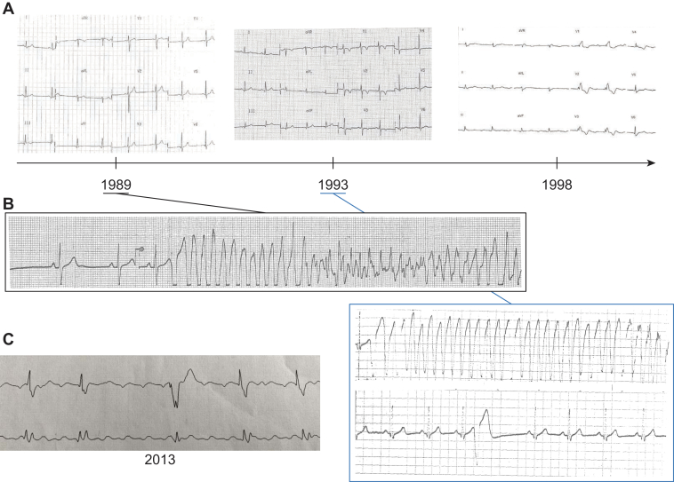
Electrocardiograms and electrical characteristics. A: Twelve-lead electrocardiograms performed at the time of the initial event (left), 4 years later (middle), and 9 years later (right). B: Short premature ventricular beats that triggered ventricular fibrillation in 1989 and 1993. C: Plot shows how the durations of the corrected QT (ms, black, left axis) and QRS (ms, red, right axis) evolved in the index patient.
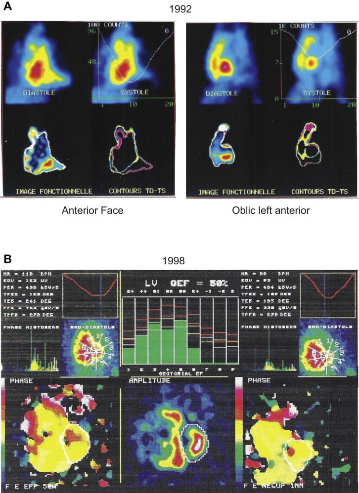
SPECT phase analysis imaging. A: Image acquired at the time of ventricular fibrillation occurrence, with no abnormalities. B: Nine years later, with frank right ventricular dilation.
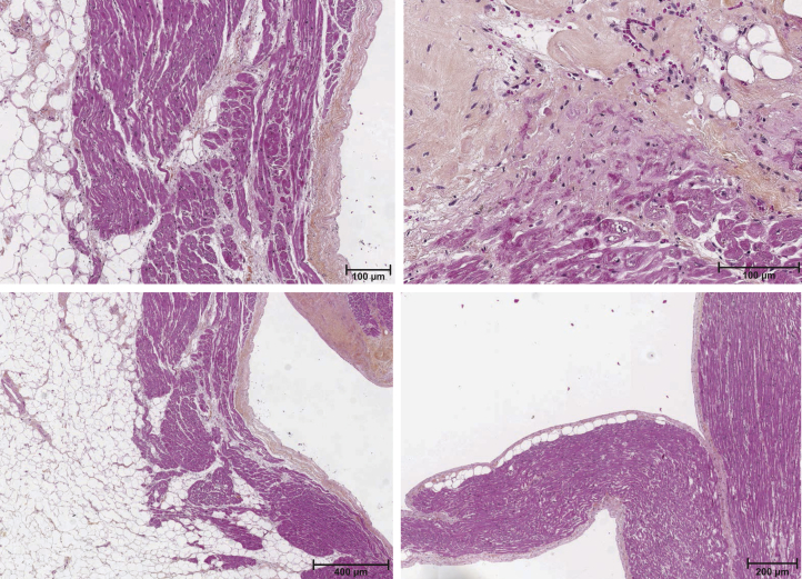
Patient’s heart autopsy. Examples of a microscopic section of the patient’s explanted heart stained with hematoxylin, eosin, and saffron. Inflammation, adipose tissue, and myocardial necrosis are observed.
