

Ureteral fibroepithelial polyp: A diagnostic challenge
- PMID: 34820289
- PMCID: PMC8602012
- DOI: 10.1016/j.eucr.2021.101940
Ureteral fibroepithelial polyp: A diagnostic challenge
Abstract
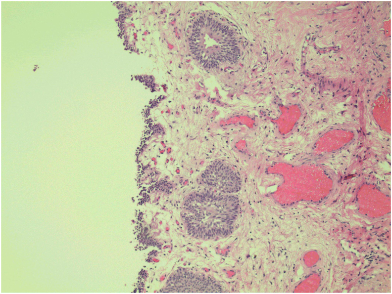
Ureteral fibroepithelial polyps (UFP) are relatively rare, benign tumors. A total of 236 total cases were documented between 1932 and 2013. Notably, imaging studies, including computerized tomography (CT) and magnetic resonance imaging (MRI) are often negative. This report details a case of a patient with a UFP who presented with hematuria. CT suggested a possible 1.8 cm mass, but subsequent MRI was negative. Cystoscopy showed a polyp that prolapsed out of the left ureter and into the bladder with peristalsis. The patient subsequently underwent retrograde ureteroscopy and holmium laser excision of the polyp.
Keywords: Hematuria; Ureteral fibroepithelial polyp; Ureteroscopy.
© 2021 The Authors.
Figures


References
-
- Liddell R.M., Weinberger E., Schofield D.E., Pelman R.S. Fibroepithelial polyp of the ureter in a child. AJR Am J Roentgenol. 1991;157(6):1273–1274. - PubMed
-
- Nowak M.A., Marzich C.S., Scheetz K.L., McElroy J.B. Benign fibroepithelial polyps of the renal pelvis. Arch Pathol Lab Med. 1999;123(9):850–852. - PubMed
-
- Lam J.S., Bingham J.B., Gupta M. Endoscopic treatment of fibroepithelial polyps of the renal pelvis and ureter. Urology. 2003;62(5):810–813. - PubMed
Publication types
LinkOut - more resources
Full Text Sources
