Review
. 2022 Jan;305(1):152-164.
doi: 10.1111/imr.13045. Epub 2021 Nov 24.

Micro but mighty-Micronutrients in the epigenetic regulation of adaptive immune responses

Affiliations
Review

Micro but mighty-Micronutrients in the epigenetic regulation of adaptive immune responses

Heng-Yi Chen et al. Immunol Rev. 2022 Jan.

Abstract

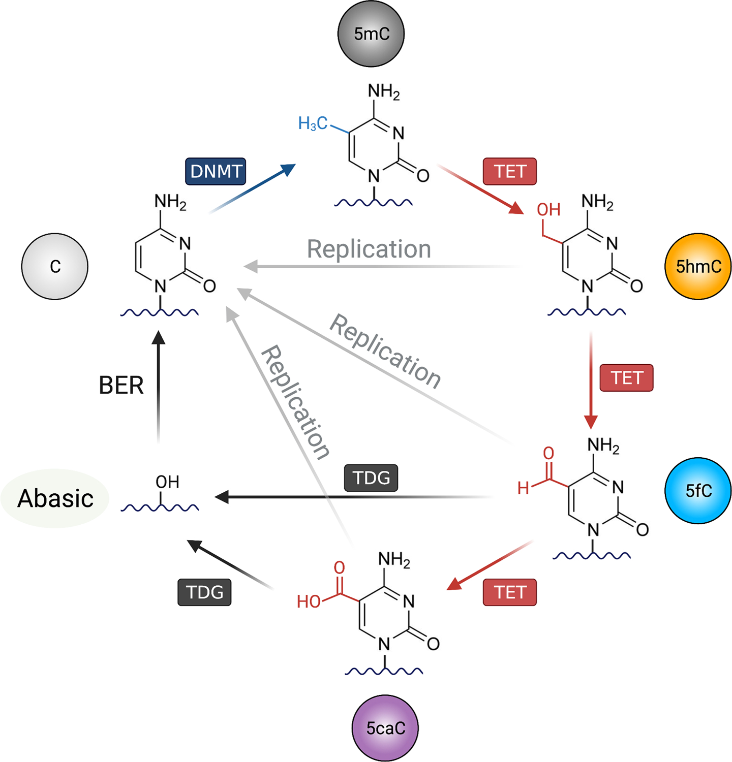
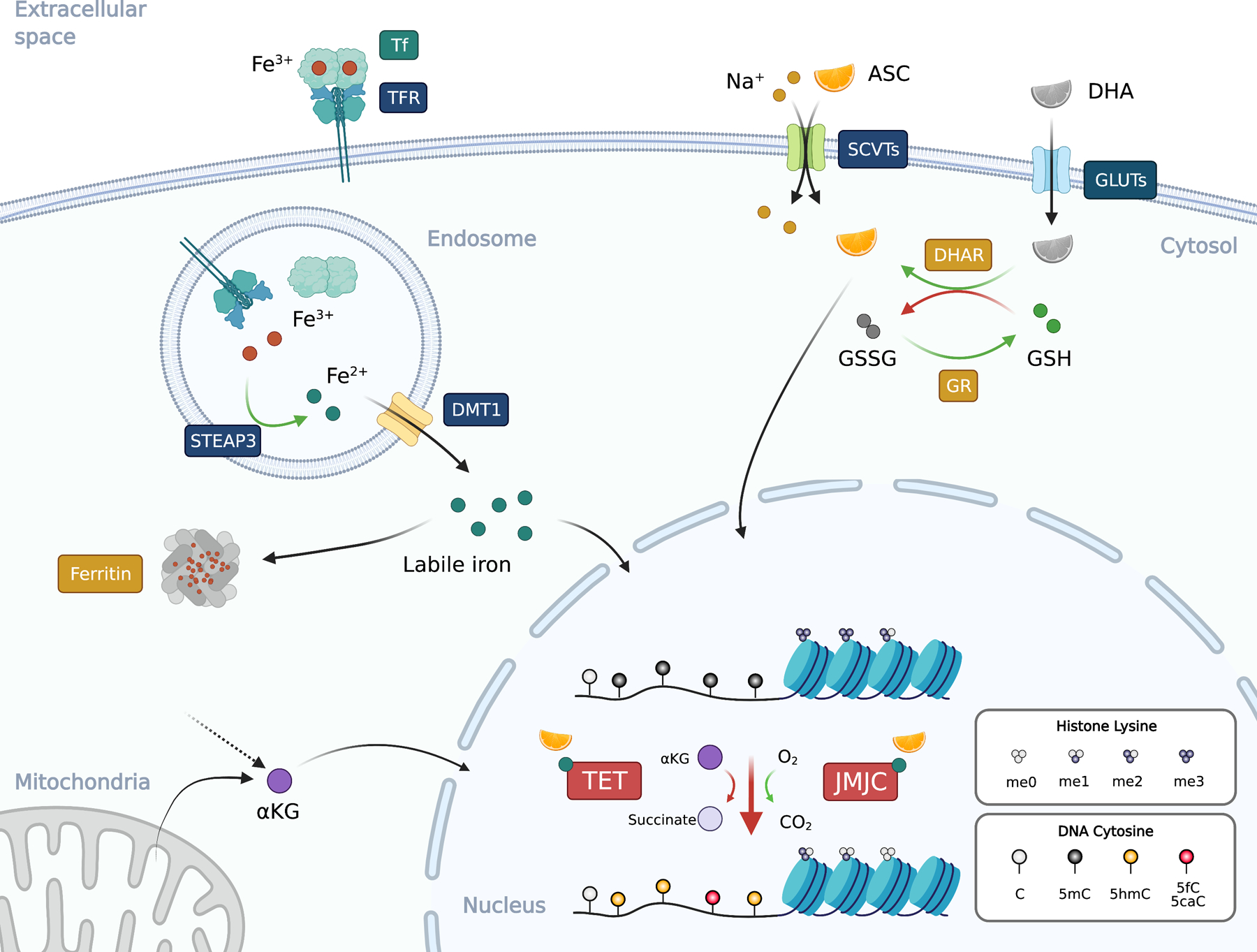
Micronutrients are essential small molecules required by organisms in minute quantity for survival. For instance, vitamins and minerals, the two major categories of micronutrients, are central for biological processes such as metabolism, cell replication, differentiation, and immune response. Studies estimated that around two billion humans worldwide suffer from micronutrient deficiencies, also known as "hidden hunger," linked to weakened immune responses. While micronutrients affect the immune system at multiple levels, recent studies showed that micronutrients potentially impact the differentiation and function of immune cells as cofactors for epigenetic enzymes, including the 2-oxoglutarate-dependent dioxygenase (2OGDD) family involved in histone and DNA demethylation. Here, we will first provide an overview of the role of DNA methylation in T cells and B cells, followed by the micronutrients ascorbate (vitamin C) and iron, two critical cofactors for 2OGDD. We will discuss the emerging evidence of these micronutrients could regulate adaptive immune response by influencing epigenetic remodeling.

Keywords: B cells; DNA methylation; T cells; epigenetics; iron; micronutrients; vitamin C.


Figures

Figure 1.
Figure 1.. The DNA methylation-demethylation cycle.
Unmodified cytosine (C) on the DNA can be methylated by the DNA methyltransferases (DNMTs), forming 5-methylcytosine (5mC). For DNA demethylation, TET (Ten-Eleven Translocation) oxidizes 5mC into oxidized cytosines including 5-hydroxymethylcytosine (5hmC), 5-formylcytosine (5fC), and 5-carboxylcytosine (5caC). There are mainly two mechanisms for TET-mediated DNA demethylation: replication-independent (active) and replication-dependent (passive) pathways. In active DNA demethylation, 5fC and 5caC are recognized and excised by thymine DNA glycosylase (TDG), creating an abasic site that is replaced with unmethylated cytosine by base excision repair (BER) pathway. In passive demethylation, while the DNMT1/UHRF1 complex recognize hemi-methylated DNA CpG on replicating DNA, the complex is not able to recognize and methylate the hemi-methylated CpG containing oxidized cytosines (5fC, 5fC and 5caC), thus inhibiting the methylation of the CpG motif on the newly replicated DNA. Figure was created with BioRender.
Figure 2.
Figure 2.. Ascorbate and iron in epigenetic regulation.
Ascorbate (ASC; vitamin C or VC) is co-transported with sodium ion (Na+) from extracellular space to the cytosol via SVCTs (sodium-ascorbate co-transporters) at the ratio of 1-to-2 (ASC to sodium). Dehydroascorbate (DHA), the oxidized form of ASC, enters the cells via the glucose transporters (GLUTs). DHA in the cells is reduced to ASC by dehydroascorbate reductase (DHAR) by oxidizing two molecules of reduced (GSH) into oxidized glutathione (GSSG). GSSG can be reduced back to GSH by glutathione reductase (GR). Iron usually exists in the circulation as the oxidized ferric iron (Fe3+) bound to transferrin (Tf) at a ratio of 2:1. Transferrin receptor (TFR) binds to and internalize the Tf:iron complex to endosome. The ferric iron is released from Tf in the acidic environment and reduced by the metalloreductase STEAP3 (Six-Transmembrane Epithelial Antigen of Prostate 3) into ferrous iron (Fe2+), which then translocates from the endosome to cytosol via DMT1 (Divalent Metal Transporter 1) to form the labile iron pool. Since ferrous iron can catalyze the formation of ROS, its concentration is tightly regulated and is stored as ferric iron in the ferritin complex that consists of 24 subunits. The ferritin complex can hold around 4,500 molecules of Fe3+. Both ASC and ferrous iron are cofactors for 2OGDDs including TET and JMJC proteins that used αKG (2-OG) and molecular oxygen to oxidize the target. TET oxidizes 5mC into 5hmC, 5fC, and 5caC, while various JMJC proteins demethylate histone lysine (and arginine, not depicted). αKG can be derived from mitochondria or cytosol as an intermediate metabolite. Black arrows, movement of molecules; green arrows, reduction; red arrows, oxidation. Blue text boxes represent transmembrane proteins and boxes with other colors indicate the location of the proteins. Figure was created with BioRender.

References

    1. In: Howson CP, Kennedy ET, Horwitz A, eds. Prevention of Micronutrient Deficiencies: Tools for Policymakers and Public Health Workers. Washington (DC), 1998. - PubMed
    1. Carr AC, Rowe S. Factors Affecting Vitamin C Status and Prevalence of Deficiency: A Global Health Perspective. Nutrients.2020;12. - PMC - PubMed
    1. Lio CJ, Huang SC. Circles of Life: linking metabolic and epigenetic cycles to immunity. Immunology.2020;161:165–174. - PMC - PubMed
    1. Dai Z, Ramesh V, Locasale JW. The evolving metabolic landscape of chromatin biology and epigenetics. Nature reviews Genetics.2020;21:737–753. - PMC - PubMed
    1. Izzo LT, Affronti HC, Wellen KE. The Bidirectional Relationship Between Cancer Epigenetics and Metabolism. Annu Rev Cancer Biol.2021;5:235–257. - PMC - PubMed

Publication types