. 2022 Jan;51(1):103-111.
doi: 10.1111/ahe.12769. Epub 2021 Nov 24.

Morphological characteristics of the blackspot seabream (Pagellus bogaraveo) tongue: A structural and immunohistochemical study

Affiliations

Morphological characteristics of the blackspot seabream (Pagellus bogaraveo) tongue: A structural and immunohistochemical study

Francesco Abbate et al. Anat Histol Embryol. 2022 Jan.

Abstract

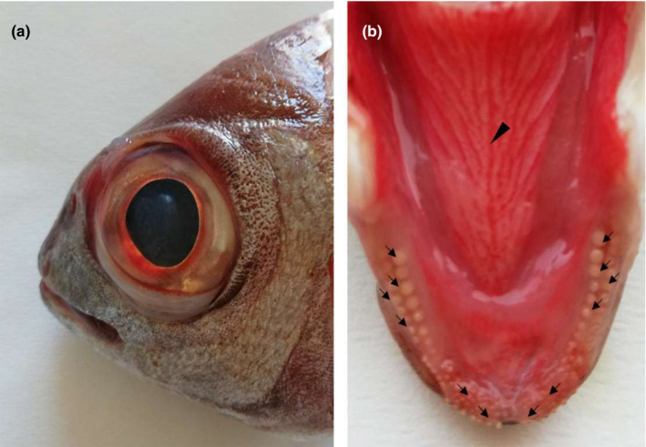
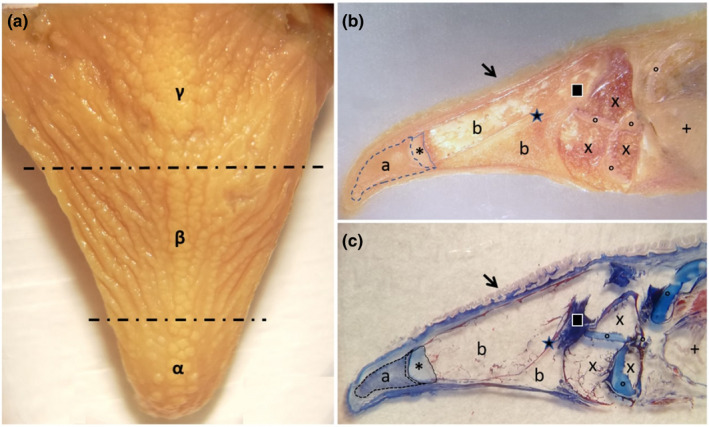
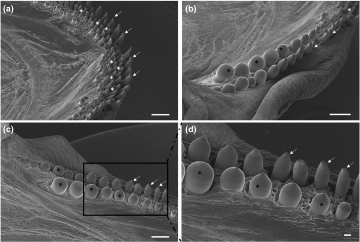
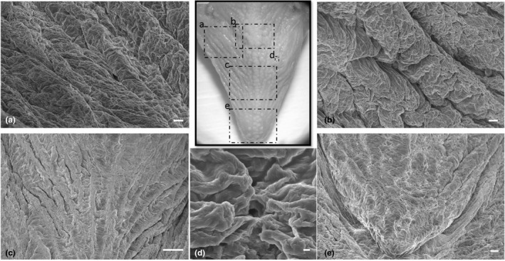
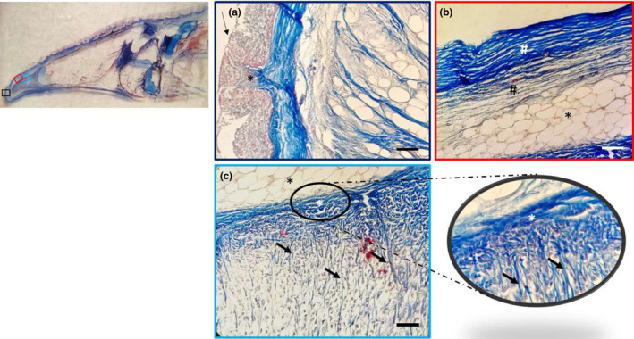
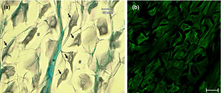
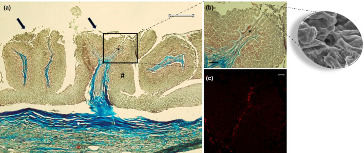
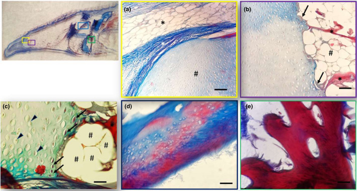
The blackspot seabream (Pagellus bogaraveo, Brünnich, 1768) is an omnivorous, predominantly carnivorous fish. In aquaculture, it is fed with pellets rich in proteins and fat. The morphological and functional aspects of the fish tongue, the feeding modality and the tasting capacity are strictly related. Therefore, the aim of this study was to describe by scanning electron, light and confocal laser microscopy, the morphological characteristics of the tongue in this species. It showed an apex, a body and a root. There were rows of teeth on the edges of the mouth and taste pores on all the tongue dorsal surface with folds and furrows. In addition, body and root showed several fungiform-like papillae in the mucosa of the folds, covered by a weakly keratinized stratified squamous epithelium, can be observed. The papillae were innervated by S100 positive fibres. In the apex, a mesenchymal tissue with vimentin positive star-shaped stem cells was evident. The results could give a support for a wider use of the blackspot seabream as a farmed species, considering the morphological data as correlated with the potentiality of food discrimination. This provides a basis for possible applications in feeding strategies. The presence, localization and characteristics of the mesenchymal stem cells, as seen also in previous studies, could represent a further basis for future applications in clinical trials.

Keywords: fish; immunohistochemistry; light microscopy; scanning electron microscopy; tongue.


Conflict of interest statement

The authors declare that they have no conflict of interest.

Figures

FIGURE 1
FIGURE 1
Gross photograph showing (a) macroscopical aspect of the blackspot seabream head and (b) the tongue dorsal surface: The arrows indicate teeth of various shapes, arranged in rows. The arrowhead indicates the median raphe
FIGURE 2
FIGURE 2
Gross photograph showing (a) macroscopical aspect of the blackspot seabream tongue, which is divided in three areas: the apex (α), the body (β) and the root (γ). Stereomicrographs (b and c) sagittal sections of the tongue: The arrow indicates the tongue dorsal surface covered by several papillae. At the apex level, a pad of mesenchimal tissue is evident (a), with aborally a cartilagineous tissue (asterisk). From the aboral portion of cartilagineous tissue, bone trabeculae were evident, originated by indirect ossification, delimitating niches of adipose tissue (b). An area of compact bone tissue (star) originates through a process of indirect ossification to replace a pre‐existing cartilagineous tissue (square). Adipose tissue (×), areas of hyaline cartilage (circles) and gills (+) characterize the aboral areas of the tongue stratigraphy
FIGURE 3
FIGURE 3
(a) Scanning electron micrograph of the rostral part of the oral cavity with cardiform (white asterisks) and caniniform teeth with aborally curved tips (arrows). (b and c) The molariphorm (black asterisks), caniniform (arrows) and cardiform (white asterisk) teeth on the oral cavity margins. (d) At higher magnification, the molariform (asterisks) and caniniform (arrows) teeth. Scale bar: a, b, c: 1mm; d: 200 μm
FIGURE 4
FIGURE 4
(a–c,). Scanning electron micrograph of the irregular dorsal surface of the tongue with prominent folds and deep furrows, with a latero‐medial orientation fading towards the apex. (d) Taste pores were observed among the folds. (e) The dorsal surface of the apex with less prominent folds. Scale bar: a, b: 100 μm; c: 1mm; d: 10 μm; e: 200 μm
FIGURE 5
FIGURE 5
Light micrographs (Masson's trichrome with aniline blue staining): (a) In the apex, a weakly keratinized stratified squamous epithelium (arrow) and connective papillae (asterisk), showing between the epithelial laminae. (b) The dense fibrillar connective tissue of the papillae with abundant collagen fibres (white hashtag) and the deeper loose fibrillar connective tissue (black hashtag), which continued with a pad of unilocular adipose tissue (asterisk). (c) Unilocular adipose tissue (black asterisk); the dense connective tissue (white asterisk) forms septa (arrows) outlining niches of cells, at higher magnification in the insert. Scale bar: 20 μm
FIGURE 6
FIGURE 6
Light micrographs (Masson's trichrome with aniline blue staining) showing (a) the mesenchymal tissue with star‐shaped cells (arrows) immersed in abundant extracellular matrix (hashtag), with an evident septum of connective tissue (asterisk). (b) Vimentin positive star‐shaped cells. Scale bar: a: 9 μm; b: 20 μm
FIGURE 7
FIGURE 7
Light micrographs (Masson's trichrome with aniline blue staining) showing (a) in the body and root fungiform‐like papillae (arrows). The mucosa (hashtag) raises in plicae showing the papillae. The connective tissue (white asterisk) throws itself in the mucosa bringing vessels and nerves (black asterisk). (b) A taste pore showed also in the scanning electron micrograph insert, and vessels and nerve (asterisk). (c) Note the presence of papillae with S100 positive nervous fibres. Scale bar: a: 200 μm; b: 30 μm; c: 20 μm
FIGURE 8
FIGURE 8
Light micrographs (Masson's trichrome with aniline blue staining) showing (a) different areas of the tongue: (a) Between the apex and the body the unilocular adipose tissue (black asterisk), the dense connective tissue (white asterisk) and the hyaline cartilage pad (hashtag). (b) Hyaline cartilagineous tissue (×) and an ossification process of the cartilage (arrows). A bone trabecula is indicated by the asterisk with a close adipose tissue (hashtags). (c) Chondrocytes (arrowheads) and degenerated chondrocytes (arrows). The bone tissue (asterisk) with a close adipose tissue (hashtags). (d) Hyaline cartilage is stained with blue and cartilage in red during the process of calcification. (e) Bone trabecula at higher magnification. Scale bar: a, b, d: 20 μm; c, e: 30 μm

References

    1. Abbate, F. , Germana, G. P , De Carlos, F. , Montalbano, G. , Laura, R. , Levanti, M. B , & Germana, A. (2006). The oral cavity of the adult zebrafish (Danio rerio). Anatomia Histologia Embryologia, 35, 299–304. 10.1111/j.1439-0264.2006.00682.x - DOI - PubMed
    1. Abbate, F. , Guerrera, M. C. , Cavallaro, M. , Montalbano, G. , Germanà, A. , & Levanti, M. (2017). LM and SEM study on the swordfish (Xiphias gladius) tongue. Tissue and Cell, 49(6), 633–637. 10.1016/j.tice.2017.09.007 - DOI - PubMed
    1. Abbate, F. , Guerrera, M. C. , Levanti, M. , Laurà, R. , Aragona, M. , Mhalhel, K. , Montalbano, G. , & Germanà, A. (2020b) Anatomical, histological and immunohistochemical study of the tongue in the rainbow trout (Oncorhynchus mykiss). Anatomia Histologia Embryologia, 49(6), 848–858. 10.1111/ahe.12593 - DOI - PubMed
    1. Abbate, F. , Guerrera, M. C. , Levanti, M. , Laurà, R. , Montalbano, G. , Cavallaro, M. , & Germanà, A. (2020a). Morphology of the Atlantic salmon (Salmo salar) tongue. Anatomia Histologia Embryologia, 49(6):686–694. 10.1111/AHE.12563 - DOI - PubMed
    1. Abbate, F. , Guerrera, M. C. , Levanti, M. , Laurà, R. , Montalbano, G. , Cavallaro, M. , & Germanà, A. (2020c). The tongue of Leopard Gecko (Eublepharis macularius). Anatomia Histologia Embryologia, 49, 51–59. - PubMed