

Incorporation and Utility of a Responsive Ribonucleoside Analogue in Probing the Conformation of a Viral RNA Motif by Fluorescence and 19 F NMR Spectroscopy
- PMID: 34821449
- PMCID: PMC7612754
- DOI: 10.1002/cbic.202100601
Incorporation and Utility of a Responsive Ribonucleoside Analogue in Probing the Conformation of a Viral RNA Motif by Fluorescence and 19 F NMR Spectroscopy
Abstract
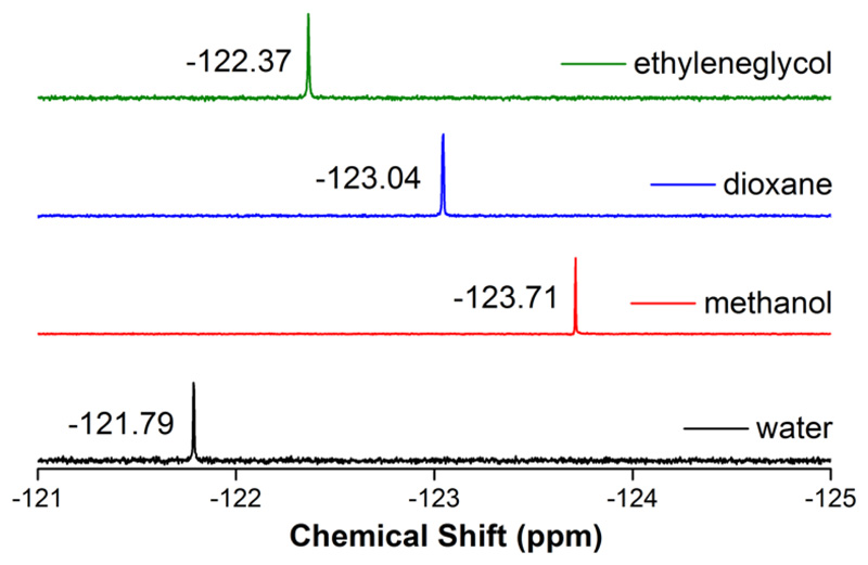
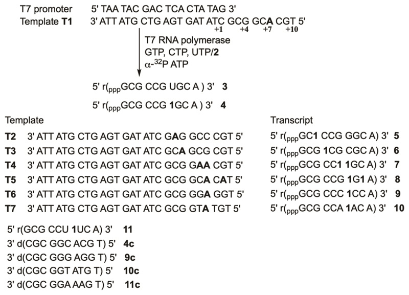
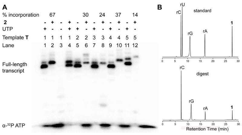
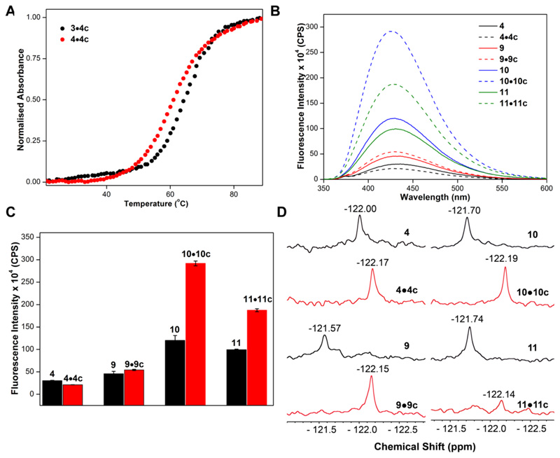
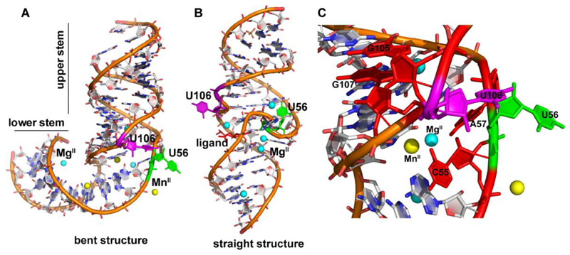
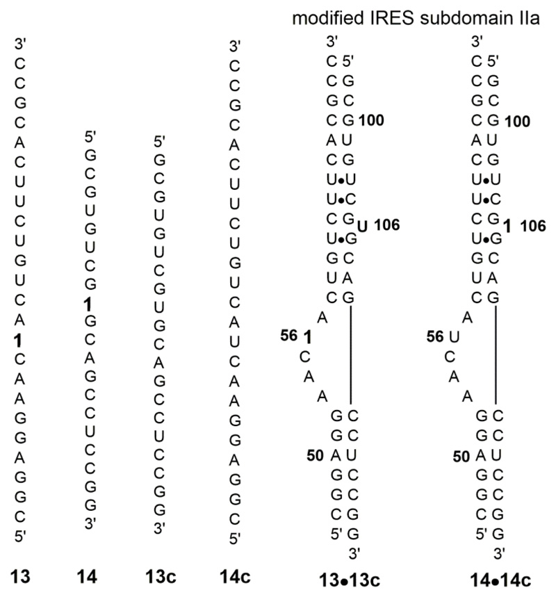
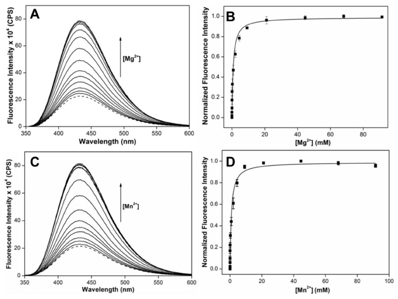
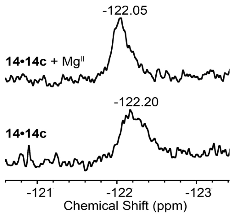
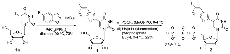
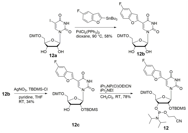
Development of versatile probes that can enable the study of different conformations and recognition properties of therapeutic nucleic acid motifs by complementing biophysical techniques can greatly aid nucleic acid analysis and therapy. Here, we report the design, synthesis and incorporation of an environment-sensitive ribonucleoside analogue, which serves as a two-channel biophysical platform to investigate RNA structure and recognition by fluorescence and 19 F NMR spectroscopy techniques. The nucleoside analogue is based on a 5-fluorobenzofuran-uracil core and its fluorescence and 19 F NMR chemical shifts are highly sensitive to changes in solvent polarity and viscosity. Notably, the modified ribonucleotide and phosphoramidite substrates can be efficiently incorporated into RNA oligonucleotides (ONs) by in vitro transcription and standard solid-phase ON synthesis protocol, respectively. Fluorescence and 19 F readouts of the nucleoside incorporated into model RNA ONs are sensitive to the neighbouring base environment. The responsiveness of the probe was aptly utilized in detecting and quantifying the metal ion-induced conformational change in an internal ribosome entry site RNA motif of hepatitis C virus, which is an important therapeutic target. Taken together, our probe is a good addition to the nucleic acid analysis toolbox with the advantage that it can be used to study nucleic acid conformation and recognition simultaneously by two biophysical techniques.
Keywords: NMR; fluorescence; nucleoside probe; ribonucleoside analogue; viral RNA.
© 2021 Wiley-VCH GmbH.
Conflict of interest statement
The authors declare no competing financial interest.
Figures

Similar articles
-
Synthesis and Enzymatic Incorporation of a Dual-App Nucleotide Probe That Reports Antibiotics-Induced Conformational Change in the Bacterial Ribosomal Decoding Site RNA.ACS Chem Biol. 2024 Mar 15;19(3):687-695. doi: 10.1021/acschembio.3c00676. Epub 2024 Feb 26. ACS Chem Biol. 2024. PMID: 38407057
-
A microenvironment-sensitive fluorescent pyrimidine ribonucleoside analogue: synthesis, enzymatic incorporation, and fluorescence detection of a DNA abasic site.Chemistry. 2011 Nov 4;17(45):12820-7. doi: 10.1002/chem.201101194. Epub 2011 Sep 28. Chemistry. 2011. PMID: 21956450
-
Site-selected introduction of modified purine and pyrimidine ribonucleosides into RNA by automated phosphoramidite chemistry.Biochimie. 1995;77(1-2):125-34. doi: 10.1016/0300-9084(96)88115-6. Biochimie. 1995. PMID: 7599270
-
Nucleoside analog inhibitors of hepatitis C virus replication.Infect Disord Drug Targets. 2006 Mar;6(1):17-29. doi: 10.2174/187152606776056698. Infect Disord Drug Targets. 2006. PMID: 16787301 Review.
-
Synthesis of 5'-O-DMT-2'-O-TBS Mononucleosides Using an Organic Catalyst.Curr Protoc Nucleic Acid Chem. 2014 Jun 24;57:2.17.1-11. doi: 10.1002/0471142700.nc0217s57. Curr Protoc Nucleic Acid Chem. 2014. PMID: 24961720 Free PMC article. Review.
References
Publication types
MeSH terms
Substances
Grants and funding
LinkOut - more resources
Full Text Sources
Miscellaneous
