The Atherosclerotic Profile of a Young Symptomatic Population between 19 and 49 Years: Coronary Computed Tomography Angiography or Coronary Artery Calcium Score?
- PMID: 34821710
- PMCID: PMC8625601
- DOI: 10.3390/jcdd8110157
The Atherosclerotic Profile of a Young Symptomatic Population between 19 and 49 Years: Coronary Computed Tomography Angiography or Coronary Artery Calcium Score?
Abstract
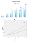
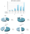
(1) Background: Whether coronary computed tomography angiography (CTA) or the coronary artery calcium score (CACS) should be used for diagnosis of coronary heart disease, is an open debate. The aim of our study was to compare the atherosclerotic profile by coronary CTA in a young symptomatic high-risk population (age, 19-49 years) in comparison with the coronary artery calcium score (CACS). (2) Methods: 1137 symptomatic high-risk patients between 19-49 years (mean age, 42.4 y) who underwent coronary CTA and CACS were stratified into six age groups. CTA-analysis included stenosis severity and high-risk-plaque criteria (3) Results: Atherosclerosis was more often detected based on CTA than based on CACS (45 vs. 27%; p < 0.001), 50% stenosis in 13.6% and high-risk plaque in 17.7%. Prevalence of atherosclerosis was low and not different between CACS and CTA in the youngest age groups (19-30 y: 5.2 and 6.4% and 30-35 y: 10.6 and 16%). In patients older than >35 years, the rate of atherosclerosis based on CTA increased (p = 0.004, OR: 2.8, 95%CI:1.45-5.89); and was higher by CTA as compared to CACS (34.9 vs. 16.7%; p < 0.001), with a superior performance of CTA. In patients older than 35 years, stenosis severity (p = 0.002) and >50% stenosis increased from 2.6 to 12.5% (p < 0.001). High-risk plaque prevalence increased from 6.4 to 26.5%. The distribution of high-risk plaque between CACS 0 and >0.1 AU was similar among all age groups, with an increasing proportion in CACS > 0.1 AU with age. A total of 24.9% of CACS 0 patients had coronary artery disease based on CTA, 4.4% > 50% stenosis and 11.5% had high-risk plaque. (4) Conclusions: In a symptomatic young high-risk population older than 35 years, CTA performed superior than CACS. In patients aged 19-35 years, the rate of atherosclerosis was similar and low based on both modalities. CACS 0 did not rule out coronary artery disease in a young high-risk population.
Keywords: atherosclerosis; computed tomography; coronary arteries; imaging; young high-risk population.
Conflict of interest statement
The authors declare no conflict of interest.
Figures


Similar articles
-
The Atherosclerosis Profile by Coronary Computed Tomography Angiography (CTA) in Symptomatic Patients with Coronary Artery Calcium Score Zero.Diagnostics (Basel). 2022 Aug 24;12(9):2042. doi: 10.3390/diagnostics12092042. Diagnostics (Basel). 2022. PMID: 36140444 Free PMC article.
-
Gender Differences in the Atherosclerosis Profile by Coronary CTA in Coronary Artery Calcium Score Zero Patients.J Clin Med. 2021 Mar 15;10(6):1220. doi: 10.3390/jcm10061220. J Clin Med. 2021. PMID: 33804095 Free PMC article.
-
Smoking and obesity predict high-risk plaque by coronary CTA in low coronary artery calcium score (CACS).J Cardiovasc Comput Tomogr. 2021 Nov-Dec;15(6):499-505. doi: 10.1016/j.jcct.2021.04.003. Epub 2021 Apr 24. J Cardiovasc Comput Tomogr. 2021. PMID: 33933380
-
Prevalence of coronary artery disease in symptomatic patients with zero coronary artery calcium score in different age population.Int J Cardiovasc Imaging. 2021 Feb;37(2):723-729. doi: 10.1007/s10554-020-02028-8. Epub 2020 Sep 26. Int J Cardiovasc Imaging. 2021. PMID: 32979114
-
Computed tomography and coronary artery calcium score for screening of coronary artery disease and cardiovascular risk management in asymptomatic individuals.Neth Heart J. 2024 Nov;32(11):371-377. doi: 10.1007/s12471-024-01897-1. Epub 2024 Oct 2. Neth Heart J. 2024. PMID: 39356452 Free PMC article. Review.
Cited by
-
Lipomatous hypertrophy of the interatrial septum: a distinct adipose tissue type in COPD?ERJ Open Res. 2024 Sep 23;10(5):00295-2024. doi: 10.1183/23120541.00295-2024. eCollection 2024 Sep. ERJ Open Res. 2024. PMID: 39319042 Free PMC article.
-
Prevalence and Clinical Correlates of Radiologically Detected Coronary Artery Disease in Chronic Obstructive Pulmonary Disease: A Cross-Sectional Observational Study.Am J Respir Crit Care Med. 2025 Jun;211(6):946-956. doi: 10.1164/rccm.202404-0838OC. Am J Respir Crit Care Med. 2025. PMID: 39680915
References
-
- Carr J.J., Jacobs D.R., Terry J.G., Shay C.M., Sidney S., Liu K., Schreiner P.J., Lewis C.E., Shikany J.M., Reis J.P., et al. Association of Coronary Artery Calcium in Adults Aged 32 to 46 Years With Incident Coronary Heart Disease and Death. JAMA Cardiol. 2017;2:391–399. doi: 10.1001/jamacardio.2016.5493. - DOI - PMC - PubMed
-
- Miedema M.D., Dardari Z.A., Nasir K., Blankstein R., Knickelbine T., Oberembt S., Shaw L., Rumberger J., Michos E.D., Rozanski A., et al. Association of Coronary Artery Calcium With Long-term, Cause-Specific Mortality Among Young Adults. JAMA Netw. Open. 2019;2:e197440. doi: 10.1001/jamanetworkopen.2019.7440. - DOI - PMC - PubMed
-
- Greenland P., Alpert J.S., Beller G.A., Benjamin E.J., Budoff M.J., Fayad Z.A., Foster E., Hlatky M.A., Hodgson J.M., Kushner F.G., et al. 2010 ACCF/AHA Guideline for Assessment of Cardiovascular Risk in Asymptomatic Adults: A Report of the American College of Cardiology Foundation/American Heart Association Task Force on Practice Guidelines Developed in Collaboration With the American Society of Echocardiography, American Society of Nuclear Cardiology, Society of Atherosclerosis Imaging and Prevention, Society for Cardiovascular Angiography and Interventions, Society of Cardiovascular Computed Tomography, and Society for Cardiovascular Magnetic Resonance. J. Am. Coll. Cardiol. 2010;56:2182–2199.
-
- Blaha M.J., Cainzos-Achirica M., Dardari Z., Blankstein R., Shaw L.J., Rozanski A., Rumberger J.A., Dzaye O., Michos E.D., Berman D.S., et al. All-cause and cause-specific mortality in individuals with zero and minimal coronary artery calcium: A long-term, competing risk analysis in the Coronary Artery Calcium Consortium. Atherosclerosis. 2019;294:72–79. doi: 10.1016/j.atherosclerosis.2019.11.008. - DOI - PMC - PubMed
-
- Ferencik M., Mayrhofer T., Bittner D.O., Emami H., Puchner S.B., Lu M.T., Meyersohn N.M., Ivanov A.V., Adami E.C., Patel M.R., et al. Use of High-Risk Coronary Atherosclerotic Plaque Detection for Risk Stratification of Patients With Stable Chest Pain: A Secondary Analysis of the PROMISE Randomized Clinical Trial. JAMA Cardiol. 2018;3:144–152. doi: 10.1001/jamacardio.2017.4973. - DOI - PMC - PubMed
LinkOut - more resources
Full Text Sources

