. 2021 Oct 23;7(11):222.
doi: 10.3390/jimaging7110222.

A Radioactive-Free Method for the Thorough Analysis of the Kinetics of Cell Cytotoxicity

Affiliations

A Radioactive-Free Method for the Thorough Analysis of the Kinetics of Cell Cytotoxicity

Claudia Coronnello et al. J Imaging. .

Abstract

The cytotoxic activity of T cells and Natural Killer cells is usually measured with the chromium release assay (CRA), which involves the use of 51Chromium (51Cr), a radioactive substance dangerous to the operator and expensive to handle and dismiss. The accuracy of the measurements depends on how well the target cells incorporate 51Cr during labelling which, in turn, depends on cellular division. Due to bystander metabolism, the target cells spontaneously release 51Cr, producing a high background noise. Alternative radioactive-free methods have been developed. Here, we compare a bioluminescence (BLI)-based and a carboxyfluorescein succinimidyl ester (CFSE)-based cytotoxicity assay to the standard radioactive CRA. In the first assay, the target cells stably express the enzyme luciferase, and vitality is measured by photon emission upon the addition of the substrate d-luciferin. In the second one, the target cells are labelled with CFSE, and the signal is detected by Flow Cytometry. We used these two protocols to measure cytotoxicity induced by treatment with NK cells. The cytotoxicity of NK cells was determined by adding increasing doses of human NK cells. The results obtained with the BLI method were consistent with those obtained with the CRA- or CFSE-based assays 4 hours after adding the NK cells. Most importantly, with the BLI assay, the kinetic of NK cells' killing was thoroughly traced with multiple time point measurements, in contrast with the single time point measurement the other two methods allow, which unveiled additional information on NK cell killing pathways.

Keywords: bioluminescence imaging; chromium release assay; cytotoxic assay; fluorescence; kinetics.


Conflict of interest statement

The authors declare no conflict of interest.

Figures

Figure 1
Figure 1
Chromium Release Assay. Chromium release assay (4 h) of IL2/IL15-NK challenged with HepG2 target cells labelled overnight with 5μCi of radioactive 51Cr. The percentage of specific lysis was determined by measuring the amount of chromium released by dying cells in the supernatant, using the formula: lysis % = (mean experimental counts per minute (cpm) − mean spontaneous cpm)/(mean maximum cpm − mean spontaneous cpm)  ×  100%. Each boxplot describes the % lysis obtained with cells from five donors.
Figure 2
Figure 2
Fluorescence-based cytotoxicity assay. HepG2 target cells were labelled with CFSE 5 µM and co-cultured with decreasing ratios, from 40:1 to 0:1, with effector IL2/IL15-NK cells for 4 h; cell killing was determined by flow cytometry. (A) Gating strategy. NK cells were marked as CD56+, target HepG2 cells were CFSE+. On an FSC/SSC plot gated on CD56+ and CFSE+, the two subpopulations were morphologically distinct. HepG2 (gated on P1) expressed 7AAD. By contrast, NK cells (gated on P3) were 7AAD-negative. Contour plots show one representative experiment out of three, with NK cells added to HepG2 at a 2.5:1 ratio. (B) Cumulative plot. At 4 h, the % of CFSE+7AAD+ dead target cells was determined by flow cytometry. The experiment was repeated three times using NK cells from different donors, and the % of dead target cells was plotted in floating boxes, where the median is indicated.
Figure 3
Figure 3
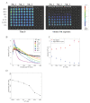
BLI-based cytotoxicity assay. In vitro quantitation of the bioluminescence signal in human hepatocellular carcinoma cells expressing luciferase, HepG2-Red-FLuc. (A) Pseudocolor representation at time 0 and after 4 h of the bioluminescence intensity of HepG2-Red-FLuc plated at 5 × 10⁴/well in a cytotoxicity assay where human NK cells from three different patients (NK_1, NK_2, and NK_3), activated with IL2/IL15, were added at decreasing ratios ranging from 40:1 to 1.25:1. (B) the thorough kinetic of cytotoxicity was addressed by measuring photon emission every two minutes for the first hour and subsequently every 5 min up to 4 h of total incubation. Average signal values and standard errors were plotted against time post NK cell incubation. (C) Peak signal and signal 4 h after NK injection plotted against NK concentration. (D) Intersection time, i.e., the time at which the time–intensity curve intersects the control time–intensity curve, plotted against NK concentration. (AD) Data shown are representative of three experiments performed in similar conditions using NK cells isolated from three different donors.

References

    1. Uzhachenko R.V., Shanker A. CD8(+) T Lymphocyte and NK Cell Network: Circuitry in the Cytotoxic Domain of Immunity. Front. Immunol. 2019;10:1906. doi: 10.3389/fimmu.2019.01906. - DOI - PMC - PubMed
    1. Timonen T., Saksela E. A simplified isotope release assay for cell-mediated cytotoxicity against anchorage dependent target cells. J. Immunol. Methods. 1977;18:123–132. doi: 10.1016/0022-1759(77)90163-6. - DOI - PubMed
    1. Nakagawa Y., Watari E., Shimizu M., Takahashi H. One-step simple assay to determine antigen-specific cytotoxic activities by single-color flow cytometry. Biomed. Res. 2011;32:159–166. doi: 10.2220/biomedres.32.159. - DOI - PubMed
    1. Gratzner H.G. Monoclonal antibody to 5-bromo- and 5-iododeoxyuridine: A new reagent for detection of DNA replication. Science. 1982;218:474–475. doi: 10.1126/science.7123245. - DOI - PubMed
    1. Fischer K., Mackensen A. The flow cytometric PKH-26 assay for the determination of T-cell mediated cytotoxic activity. Methods. 2003;31:135–142. doi: 10.1016/S1046-2023(03)00123-3. - DOI - PubMed

LinkOut - more resources