. 2021 Oct 29;7(11):229.
doi: 10.3390/jimaging7110229.

X-ray Imaging Investigation on the Gilding Technique of an Ancient Egyptian Taweret Wooden Statuette

Affiliations

X-ray Imaging Investigation on the Gilding Technique of an Ancient Egyptian Taweret Wooden Statuette

Luisa Vigorelli et al. J Imaging. .

Abstract

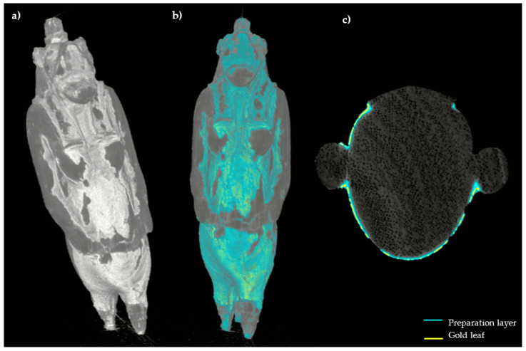
Diagnostic physical methods are increasingly applied to Cultural Heritage both for scientific investigations and conservation purposes. In particular, the X-ray imaging techniques of computed tomography (CT) and digital radiography (DR) are non-destructive investigation methods to study an object, being able to give information on its inner structure. In this paper, we present the results of the X-ray imaging study on an ancient Egyptian statuette (Late Period 722-30 BCE) belonging to the collection of Museo Egizio in Torino and representing an Egyptian goddess called Taweret, carved on wood and gilded with some colored details. Since few specific studies have been focused on materials and techniques used in Ancient Egypt for gilding, a detailed investigation was started in order to verify the technical features of the decoration in this sculpture. Specifically, DR and CT analyses have been performed at the Centro Conservazione e Restauro "La Venaria Reale" (CCR), with a new high resolution flat-panel detector, that allowed us to perform tomographic analysis reaching a final resolution better than the one achievable with the previous apparatus operating in the CCR.

Keywords: ancient Egypt; archaeometry; conservation; cultural heritage; gilding; tomography; wooden sculpture.


Conflict of interest statement

The authors declare no conflict of interest.

Figures

Figure 1
Figure 1
The analyzed Taweret statuette (Cat. 528), frontal (a) and lateral (b) views, before the conservation treatment.
Figure 2
Figure 2
Some details of the statuette: (a) face and notches for the wig (yellow arrows); (b) detail of the body where the wood material (green arrow) and the preparation layer (blue arrow) are visible.
Figure 3
Figure 3
Radiographs of the statuette, (a) frontal and (b) lateral view; (c) detail of the legs and tail insertion in the modern base.
Figure 4
Figure 4
CT slices of the statuette: (a,b) horizontal sections; (ce) longitudinal slices. In all the sections the wood structure (vessels and growing rings) is visible (green arrows: preparation layer in the face area; blue arrows: fractures inside the preparation layer; orange arrows: thin preparation material for the gold leaf adhesion).
Figure 5
Figure 5
CT longitudinal (a) and horizontal (b,c) slices (yellow arrows: wing grooves; blue arrow: fractures inside the preparation layer; pink arrow: material layer above the gold leaf).
Figure 6
Figure 6
(a) Enlargement of Figure 5c and (b) the profile plots along A and B (green arrow: preparation layer; orange arrow: preparation material for the gold leaf adhesion; pink arrow: material layer above the gold leaf).
Figure 7
Figure 7
CT horizontal slices of the body (a) and the legs (b) (orange arrow: thin preparation material for the gold leaf adhesion; purple arrow: thick preparation material between the legs).
Figure 8
Figure 8
(a) Backscattering image by means of SEM, the gold layer is in light gray; (b) enlargement of Figure 5c; (c) profile plot along A line shown in (b), only wood; (d) profile plot along B line shown in (b), gold on thin preparation layer.
Figure 9
Figure 9
3D model of the statuette with different surface render tools, ((a), Scatter Gradient v1.2 tool), ((b), Hardware render (scatter gradient) tool); enlargement of the body area in which the cracking is visible (yellow arrows) (c).
Figure 10
Figure 10
3D model of the statuette ((a), Hardware render (scatter gradient) tool) with the segmentation performed on the surface (b); CT horizontal slice in which the segmentation results are highlighted (c).

References

    1. Lang J., Middleton A.A. Radiography of Cultural Material. 2nd ed. Elsevier Butterworth-Heinemann; Oxford, UK: 2005.
    1. Casali F. X-ray and Neutron Digital Radiography and Computed Tomography for Cultural Heritage Physical Techniques in the Study of Art, Archaeology and Cultural Heritage. Elsevier; Amsterdam, The Netherlands: 2006. pp. 41–123.
    1. Morigi M.P., Casali F., Bettuzzi M., Brancaccio R., D’Errico V. Application of X-ray Computed Tomography to Cultural Heritage diagnostics. Appl. Phys. A. 2010;100:653–661. doi: 10.1007/s00339-010-5648-6. - DOI
    1. Conroy G.C., Vannier M.W. Noninvasive three-dimensional computer imaging of matrix-filled fossil skulls by high resolution computed tomography. Science. 1984;226:456–458. doi: 10.1126/science.226.4673.456. - DOI - PubMed
    1. Wind J. Computerized X-ray tomography of fossil hominid skulls. Am. J. Phys. Anthropol. 1984;63:265–282. doi: 10.1002/ajpa.1330630303. - DOI - PubMed

LinkOut - more resources