. 2022 Jan 7;375(6576):50-57.
doi: 10.1126/science.abm4245. Epub 2021 Nov 25.

Structural basis of branch site recognition by the human spliceosome

Affiliations

Structural basis of branch site recognition by the human spliceosome

Jonas Tholen et al. Science. .

Abstract

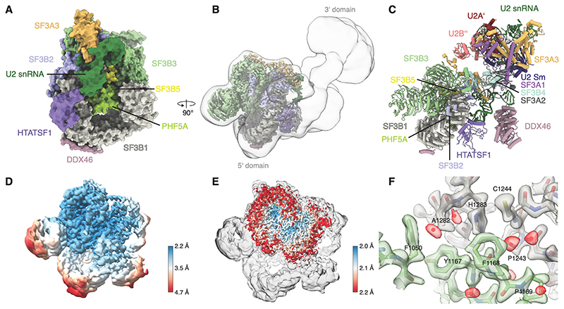
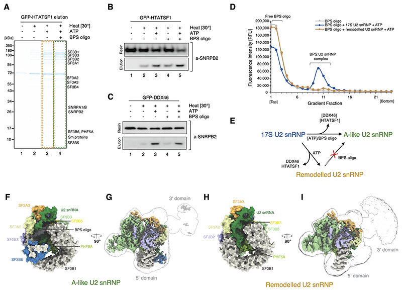
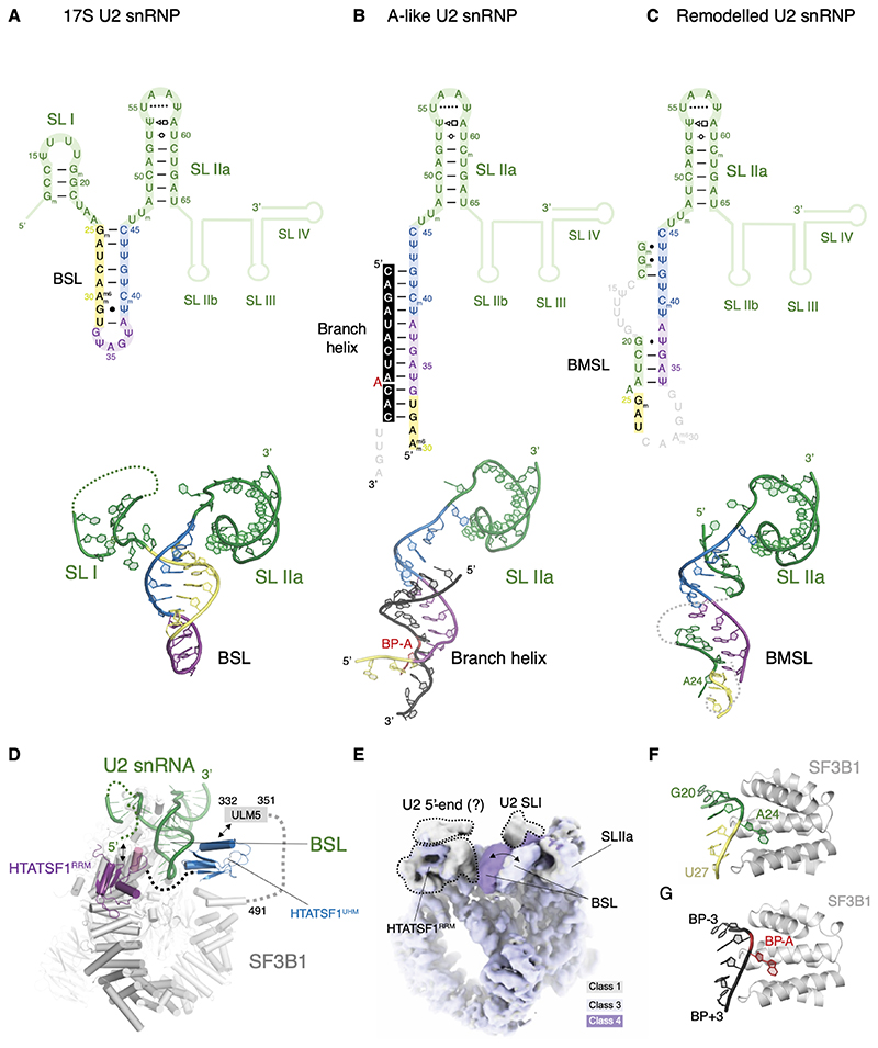
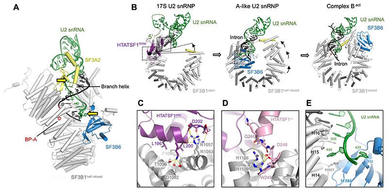
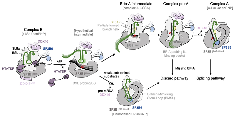
Recognition of the intron branch site (BS) by the U2 small nuclear ribonucleoprotein (snRNP) is a critical event during spliceosome assembly. In mammals, BS sequences are poorly conserved, and unambiguous intron recognition cannot be achieved solely through a base-pairing mechanism. We isolated human 17S U2 snRNP and reconstituted in vitro its adenosine 5´-triphosphate (ATP)–dependent remodeling and binding to the pre–messenger RNA substrate. We determined a series of high-resolution (2.0 to 2.2 angstrom) structures providing snapshots of the BS selection process. The substrate-bound U2 snRNP shows that SF3B6 stabilizes the BS:U2 snRNA duplex, which could aid binding of introns with poor sequence complementarity. ATP-dependent remodeling uncoupled from substrate binding captures U2 snRNA in a conformation that competes with BS recognition, providing a selection mechanism based on branch helix stability.


Conflict of interest statement

Competing interests: Authors declare that they have no competing interests.

Figures

Figure 1
Figure 1. High-resolution structure of the human 17S U2 snRNP.
(A) Surface representation of the 5’-domain of the 17S U2 snRNP model. (B) Experimental cryo-EM map for the 17S U2 snRNP showing the high-resolution 5’-domain (coloured by chain identity) embedded in a low-pass filtered map showing position of the 3’-domain. (C) Pseudo-atomic model for the fully assembled 17S U2 snRNP. 3’-domain was modelled by rigid-body docking of the previously reported coordinates (PDB:6Y5Q). (D) Cryo-EM map of the 17S U2 snRNP filtered and coloured by local resolution. (E) The cryo-EM map obtained by merging several U2 snRNP datasets overlaid with the map of the 17S U2 snRNP. (F) Atomic modelling into the highest-resolution region at an interface of SF3B1 and SF3B3. The map was coloured by chain identity, water molecules are coloured red.
Figure 2
Figure 2. Sample preparation and in vitro reconstitution of the branch site recognition by the U2 snRNP.
(A) SDS-PAGE analysis of the eluates from the GFP-HTATSF1-tagged 17S U2 snRNP immobilised on the GFP nanobody resin and incubated under various conditions. (B) Western blot analysis of the reconstitution reaction performed as in (A). Elution and resin fractions were probed with antibodies against SNRPB2, a core U2 snRNP component. (C) The same as in (B), but the 17S U2 snRNP sample was immobilised using GFP tag attached to DDX46. (D) Analysis of the Cy5-labelled BPS oligonucleotide binding to the 17S U2 snRNP or remodelled U2 snRNP by glycerol gradients. RFU, Relative Fluorescence Units. (E) Schematic summarising the outcome of the in vitro remodelling and substrate binding experiments. (F) and (H) Surface representation of the 5’-domains of the A-like and Remodelled U2 snRNPs models. (G) and (I) Experimental cryo-EM maps of A-like and Remodelled U2 snRNPs showing the high-resolution 5’-domain (coloured by chain identity) embedded in a low-pass filtered (5 Å) maps, showing positioning of their 3’-domains.
Figure 3
Figure 3. Structure and dynamics of the U2 snRNA during branch site recognition.
Secondary and tertiary structure of the U2 snRNA in the (A) 17S, (B) A-like and (C) remodelled U2 snRNPs. (D) BSL is stabilised directly by the two domains of HTATSF1 in the 17S U2 snRNP complex. (E) Structural dynamics of the BSL visualised directly in the cryo-EM map (see also movie S2). (F) Adenosine 24 of the U2 snRNA mimics BP-A in the remodelled U2 snRNP complex. (G) Environment of the BP-A in the A-like U2 snRNP in the same orientation as in (F).
Figure 4
Figure 4. SF3B6 stabilises branch helix in the A-like U2 snRNP while SF3B1 HEAT repeats adopt a half-closed conformation.
(A) Side view of the A-like U2 snRNP showing positions of the branch helix and its stabilisation by the SF3A2 and SF3B6, yellow arrows indicate U2 snRNA contact points enforcing helical geometry of the branch helix. (B) Structure of the RNA and HEAT repeats in the 17S U2 snRNP, the A-like U2 snRNP and the Bact complex (PDB: 6FF7) showing two-step transition from open to close SF3B1 conformation. (C) and (D) Atomic model of the interfaces between SF3B1HEAT and HTATSF1RRM/LH. (E) Atomic model of the interface of SF3B6 with SF3B1HEAT and U2 snRNA.
Figure 5
Figure 5. Schematic model of branch site recognition by the U2 snRNP based on recent structural data.
U2 snRNP associated with spliceosomal complex E is likely structurally similar to the 17S U2 snRNP described by (12) and in this work. Dissociation of HTATSF1 creates competition between the formation of a branch helix and the BMSL. Rejection of weak, sub-optimal substrates results in the remodelled U2 snRNP, which is targeted to a discard pathway (this work). Stable substrates gradually form the branch helix as shown in the E-to-A (41) and pre-A (42) intermediates. In the absence of properly positioned, bulged out BP-A, the pre-A complex is targeted to a discard pathway. Productive engagement of the branch helix leads to the formation of complex A, wherein U2 snRNP is structurally similar to the A-like U2 snRNP (this work).

References

    1. Valcárcel J, Gaur RK, Singh R, Green MR. Interaction of U2AF65 RS region with pre-mRNA branch point and promotion of base pairing with U2 snRNA [corrected] Science. 1996;273:1706–1709. - PubMed
    1. Michaud S, Reed R. An ATP-independent complex commits pre-mRNA to the mammalian spliceosome assembly pathway. Genes Dev. 1991;5:2534–2546. - PubMed
    1. Das R, Zhou Z, Reed R. Functional association of U2 snRNP with the ATP-independent spliceosomal complex E. Mol Cell. 2000;5:779–787. - PubMed
    1. Wu J, Manley JL. Mammalian pre-mRNA branch site selection by U2 snRNP involves base pairing. Genes Dev. 1989;3:1553–1561. - PubMed
    1. Yan D, et al. CUS2, a yeast homolog of human Tat-SF1, rescues function of misfolded U2 through an unusual RNA recognition motif. Mol Cell Biol. 1998;18:5000–5009. - PMC - PubMed

Publication types

MeSH terms