

Surface Glucan Structures in Aeromonas spp
- PMID: 34822520
- PMCID: PMC8625153
- DOI: 10.3390/md19110649
Surface Glucan Structures in Aeromonas spp
Abstract
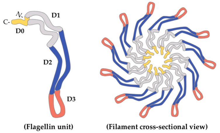
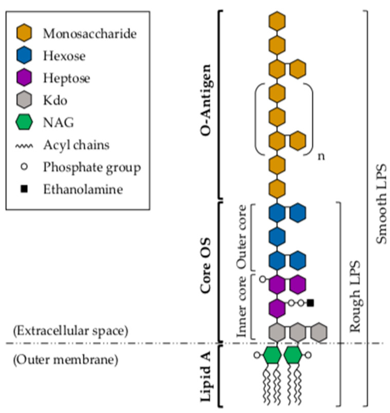
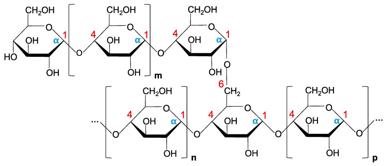
Aeromonas spp. are generally found in aquatic environments, although they have also been isolated from both fresh and processed food. These Gram-negative, rod-shaped bacteria are mostly infective to poikilothermic animals, although they are also considered opportunistic pathogens of both aquatic and terrestrial homeotherms, and some species have been associated with gastrointestinal and extraintestinal septicemic infections in humans. Among the different pathogenic factors associated with virulence, several cell-surface glucans have been shown to contribute to colonization and survival of Aeromonas pathogenic strains, in different hosts. Lipopolysaccharide (LPS), capsule and α-glucan structures, for instance, have been shown to play important roles in bacterial-host interactions related to pathogenesis, such as adherence, biofilm formation, or immune evasion. In addition, glycosylation of both polar and lateral flagella has been shown to be mandatory for flagella production and motility in different Aeromonas strains, and has also been associated with increased bacterial adhesion, biofilm formation, and induction of the host proinflammatory response. The main aspects of these structures are covered in this review.
Keywords: Aeromonas; LPS; O-antigen; capsule polysaccharide; glycosylation; α-glucan.
Conflict of interest statement
The authors declare no conflict of interest.
Figures
References
-
- Martin-Carnahan A., Joseph S.W. Order XII. Aeromonadales ord. nov. In: Brenner D.J., Krieg N.R., Staley J.T., editors. Bergey’s Manual® of Systematic Bacteriology. 2nd ed. Volume 2. Springer; Boston, MA, USA: 2005. pp. 556–587. Part B. - DOI
-
- Galindo C.L., Chopra A.K. Aeromonas and Plesiomonas species. In: Doyle M.P., Beuchat L.R., Montville T.J., editors. Food Microbiology: Fundamentals and Frontiers. 3rd ed. ASM Press; Washington, DC, USA: 2007. pp. 381–400.
-
- Joseph S.W., Carnahan A. The isolation, identification, and systematics of the motile Aeromonas species. Annu Rev. Fish. Dis. 1994;4:315–343. doi: 10.1016/0959-8030(94)90033-7. - DOI
Publication types
MeSH terms
Substances
LinkOut - more resources
Full Text Sources
Miscellaneous
