. 2021 Nov 2;9(11):289.
doi: 10.3390/toxics9110289.

Toxicity and Functional Tissue Responses of Two Freshwater Fish after Exposure to Polystyrene Microplastics

Affiliations

Toxicity and Functional Tissue Responses of Two Freshwater Fish after Exposure to Polystyrene Microplastics

Martha Kaloyianni et al. Toxics. .

Abstract

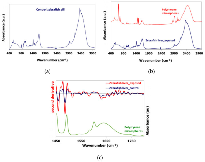
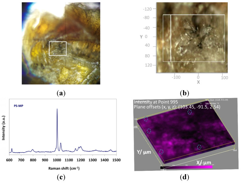
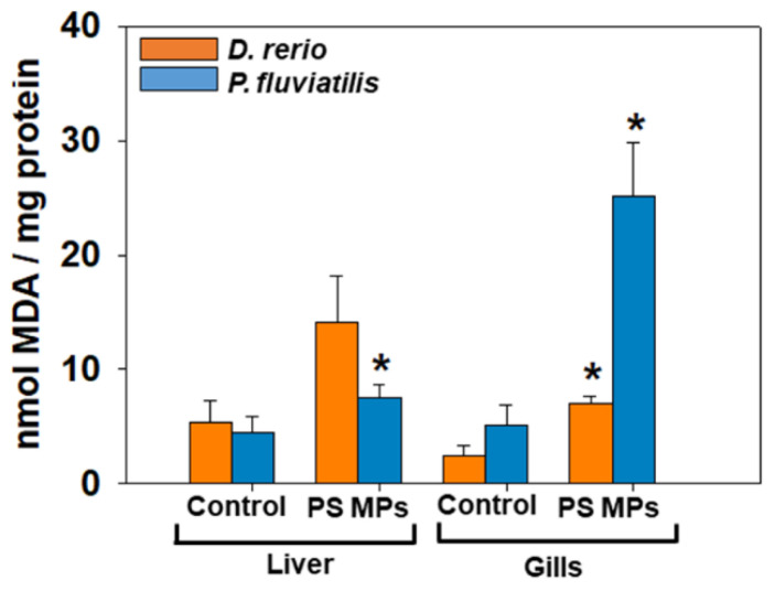
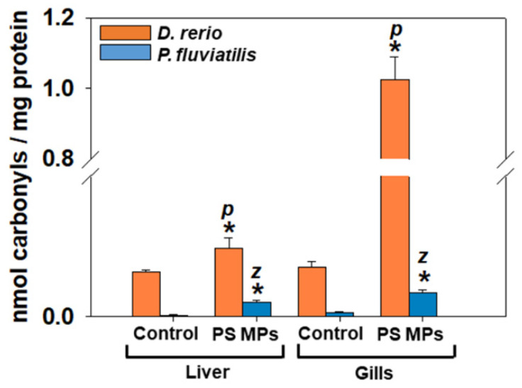
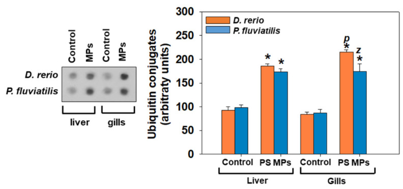
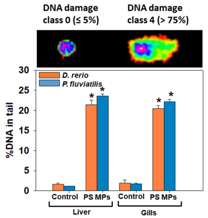
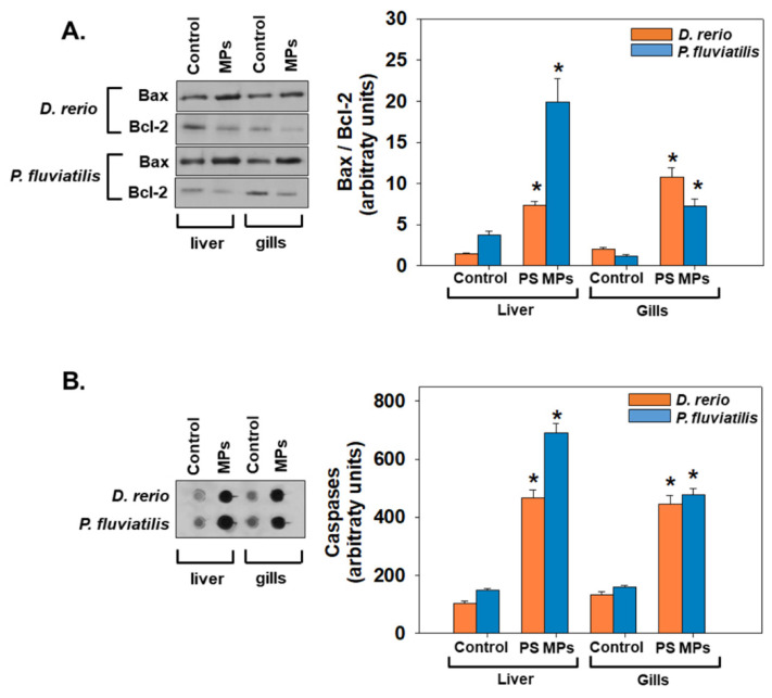
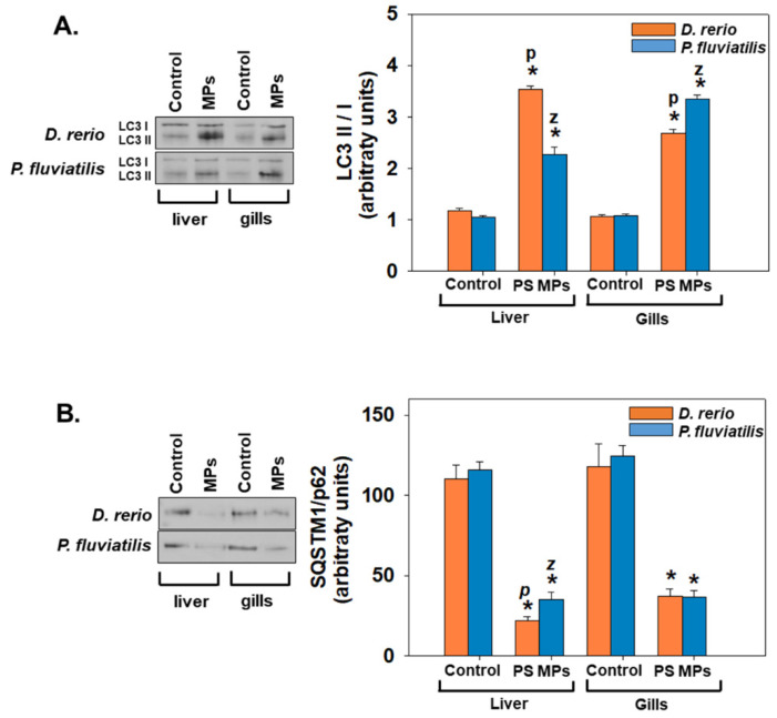
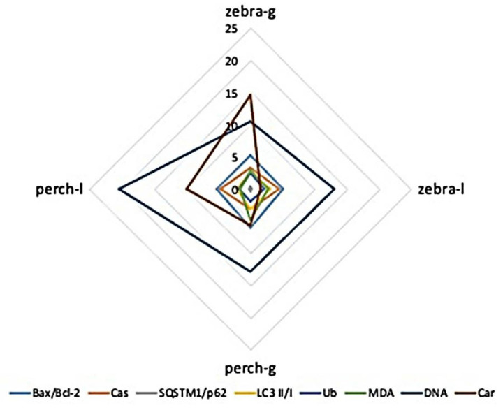
Microplastics (MPs)' ingestion has been demonstrated in several aquatic organisms. This process may facilitate the hydrophobic waterborne pollutants or chemical additives transfer to biota. In the present study the suitability of a battery of biomarkers on oxidative stress, physiology, tissue function and metabolic profile was investigated for the early detection of adverse effects of 21-day exposure to polystyrene microplastics (PS-MPs, sized 5-12 μm) in the liver and gills of zebrafish Danio rerio and perch, Perca fluviatilis, both of which are freshwater fish species. An optical volume map representation of the zebrafish gill by Raman spectroscopy depicted 5 μm diameter PS-MP dispersed in the gill tissue. Concentrations of PS-MPs close to the EC50 of each fish affected fish physiology in all tissues studied. Increased levels of biomarkers of oxidative damage in exposed fish in relation to controls were observed, as well as activation of apoptosis and autophagy processes. Malondialdehyde (MDA), protein carbonyls and DNA damage responses differed with regard to the sensitivity of each tissue of each fish. In the toxicity cascade gills seemed to be more liable to respond to PS-MPs than liver for the majority of the parameters measured. DNA damage was the most susceptible biomarker exhibiting greater response in the liver of both species. The interaction between MPs and cellular components provoked metabolic alterations in the tissues studied, affecting mainly amino acids, nitrogen and energy metabolism. Toxicity was species and tissue specific, with specific biomarkers responding differently in gills and in liver. The fish species that seemed to be more susceptible to MPs at the conditions studied, was P. fluviatilis compared to D. rerio. The current findings add to a holistic approach for the identification of small sized PS-MPs' biological effects in fish, thus aiming to provide evidence regarding PS-MPs' environmental impact on wild fish populations and food safety and adequacy.

Keywords: Danio rerio; Perca fluviatilis; gills; liver; metabolomics; microplastics; oxidative stress biomarkers; polystyrene.


Conflict of interest statement

The authors declare no conflict of interest.

Figures

Figure 1
Figure 1
(a) FTIR spectrum of the control zebrafish gill, (b) FTIR spectrum of the exposed zebrafish liver sample and the PS microspheres, (c) Second derivative analysis of the exposed and control zebrafish liver samples and the absorbance spectrum of the PS microspheres.
Figure 2
Figure 2
(a) Zebrafish gill as observed from the microscope lens, indicating the measured area with the white rectangle, (b) The measured area of Figure 2a in the x, y, z axes, (c) Raman spectrum of PS-MS, (d) Still image of the HR volume video map indicating the PS-MS in depth 2.54 μm of the gill area (the HR volume video map can be found in the Supplementary Material).
Figure 3
Figure 3
Lipid peroxidation was expressed as MDA concentrations (mean ± SD nmol mg−1 protein), in liver and gills of Danio rerio (n = 3 pools of 10 individuals) and Perca fluviatilis (n = 6). Mann–Whitney U test was employed to test for significance at p < 0.05 between all experimental groups. * denotes significant differences (p < 0.05) compared to the control group (n = 3 pools of 10 individuals and n = 6 for D. rerio and P. fluviatilis respectively).
Figure 4
Figure 4
Protein carbonylation (mean ± SD nmol carbonyls mg−1 protein) in liver and gills of Danio rerio (n = 3 pools of 10 individuals) and Perca fluviatilis (n = 6). The results were expressed as nmol carbonyl groups mg−1 of protein. Mann–Whitney U test was employed to test for significance at p < 0.05 between all experimental groups. * denotes significant differences (p < 0.05) compared to the control group (n = 3 pools of 10 individuals and n = 6 for D. rerio and P. fluviatilis respectively), while z and p denote significant differences (p < 0.05) between D. rerio and P. fluviatilis respectively.
Figure 5
Figure 5
Ubiquitin conjugates (mean ± SD) in liver and gills of Danio rerio (n = 3 pools of 10 individuals) and Perca fluviatilis (n = 6). Mann–Whitney U test was employed to test for significance at p < 0.05 between all experimental groups. * denotes significant differences (p < 0.05) compared to the control group (n = 3 pools of 10 individuals and n = 6 for D. rerio and P. fluviatilis respectively), while z and p denote significant differences (p < 0.05) between D. rerio and P. fluviatilis respectively.
Figure 6
Figure 6
Percentage (%) of DNA damage (mean ± SD) in tail in liver and gills of Danio rerio (n = 3 pools of 10 individuals) and Perca fluviatilis (n = 6). Six slides per pool (zebrafish) and six slides per individual (perch) were measured, in order to represent technical replicates. Randomly selected 100 cells were scored from each slide (TritekCometscoreTM 1.5, TriTek Corporation, Wilmington, DE, USA). Representative pictures of DNA damage are shown. Mann–Whitney U test was employed to test for significance at p < 0.05 between all experimental groups. * denotes significant differences (p < 0.05) compared to the control group (n = 3 pools of 10 individuals and n = 6 for D. rerio and P. fluviatilis respectively). % DNA in tail and Olive moment in positive control data (1 μΜ H2O2) were 28.3 ± 5.2 and 40 ± 6.3, respectively.
Figure 7
Figure 7
Bax/Bcl-2 ratio (A) and caspases levels (B) (mean ± SD) in liver and gills of Danio rerio (n = 3 pools of 10 individuals each pool) and Perca fluviatilis (n = 6). Tissue extracts from all groups were immunoblotted for Bax, Bcl-2 and caspases. Blots and dots were quantified using scanning densitometry. Representative blots and dots are shown. Mann–Whitney U test was employed to test for significance at p < 0.05 between all experimental groups. * denotes significant differences (p < 0.05) compared to the control group (n = 3 pools of 10 individuals and n = 6 for D. rerio and P. fluviatilis respectively).
Figure 8
Figure 8
LC3II/I ratio (A) and SQSTM1/p62 levels (B) (mean ± SD) in liver and gills of Danio rerio (n = 3 pools of 10 individuals) and Perca fluviatilis (n = 6). Tissue extracts from all groups were immunoblotted for LC3II/I and SQSTM1/p62. Blots and dots were quantified using scanning densitometry. Representative blots and dots are shown. Mann–Whitney U test was employed to test for significance at p < 0.05 between all experimental groups. * denotes significant differences (p < 0.05) compared to the control group (n = 3 pools of 10 individuals and n = 6 for D. rerio and P. fluviatilis respectively), while z and p denote significant differences (p < 0.05) between D. rerio and P. fluviatilis respectively.
Figure 9
Figure 9
Graphical representation of molecular and biochemical biomarkers measured in the gills (g) and liver (l) of Danio rerio (zebrafish) and Perca fluviatilis (perch) (for graphical purposes caspases and ubiquitin values were divided by 10).

References

    1. Geyer R., Jambeck J.R., Law K.L. Production, use, and fate of all plastics ever made. Sci. Adv. 2017;3:e1700782. doi: 10.1126/sciadv.1700782. - DOI - PMC - PubMed
    1. Setälä O., Fleming-Lehtinen V., Lehtiniemi M. Ingestion and transfer of microplastics in the planktonic food web. Environ. Pollut. 2014;185:77–83. doi: 10.1016/j.envpol.2013.10.013. - DOI - PubMed
    1. Cole M., Lindeque P., Fileman E., Halsband C., Goodhead R., Moger J., Galloway T.S. Microplastic Ingestion by Zooplankton. Environ. Sci. Technol. 2013;47:6646–6655. doi: 10.1021/es400663f. - DOI - PubMed
    1. Mattsson K., Ekvall M.T., Hansson L.-A., Linse S., Malmendal A., Cedervall T. Altered Behavior, Physiology, and Metabolism in Fish Exposed to Polystyrene Nanoparticles. Environ. Sci. Technol. 2015;49:553–561. doi: 10.1021/es5053655. - DOI - PubMed
    1. Della Torre C., Bergami E., Salvati A., Faleri C., Cirino P., Dawson K.A., Corsi I. Accumulation and Embryotoxicity of Polystyrene Nanoparticles at Early Stage of Development of Sea Urchin Embryos Paracentrotus lividus. Environ. Sci. Technol. 2014;48:12302–12311. doi: 10.1021/es502569w. - DOI - PubMed

LinkOut - more resources