Randomized Controlled Trial
. 2021 Nov 24;184(24):5886-5901.e22.
doi: 10.1016/j.cell.2021.11.001.

From structure to clinic: Design of a muscarinic M1 receptor agonist with potential to treatment of Alzheimer's disease

Affiliations
Randomized Controlled Trial

From structure to clinic: Design of a muscarinic M1 receptor agonist with potential to treatment of Alzheimer's disease

Alastair J H Brown et al. Cell. .

Abstract

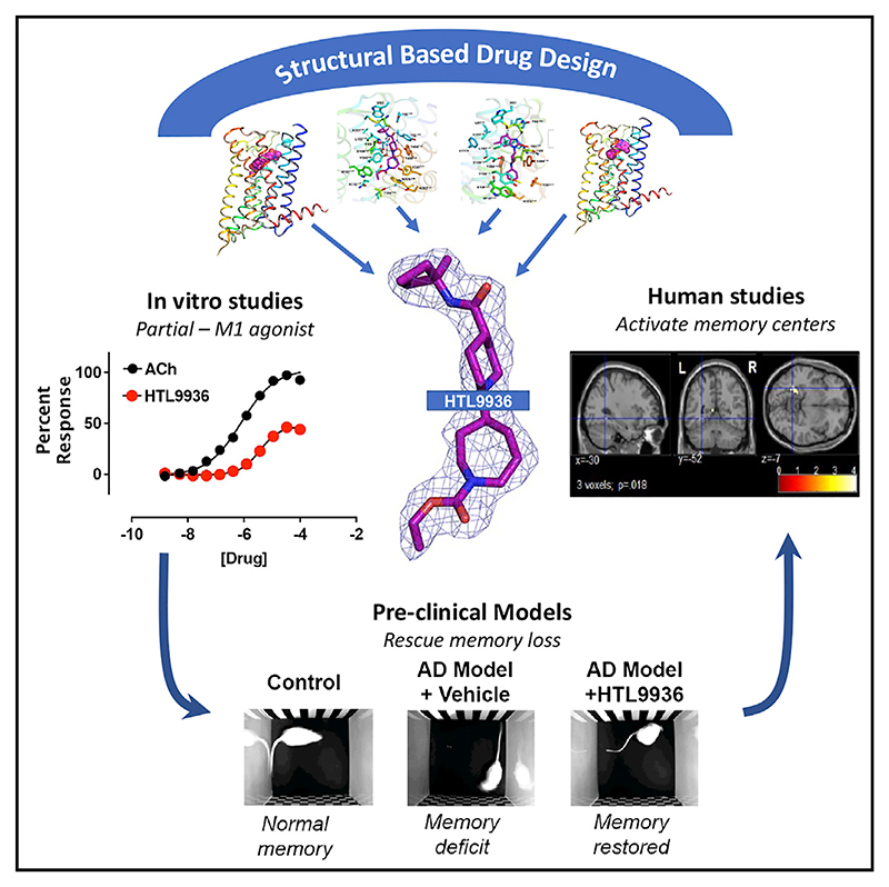
Current therapies for Alzheimer's disease seek to correct for defective cholinergic transmission by preventing the breakdown of acetylcholine through inhibition of acetylcholinesterase, these however have limited clinical efficacy. An alternative approach is to directly activate cholinergic receptors responsible for learning and memory. The M1-muscarinic acetylcholine (M1) receptor is the target of choice but has been hampered by adverse effects. Here we aimed to design the drug properties needed for a well-tolerated M1-agonist with the potential to alleviate cognitive loss by taking a stepwise translational approach from atomic structure, cell/tissue-based assays, evaluation in preclinical species, clinical safety testing, and finally establishing activity in memory centers in humans. Through this approach, we rationally designed the optimal properties, including selectivity and partial agonism, into HTL9936-a potential candidate for the treatment of memory loss in Alzheimer's disease. More broadly, this demonstrates a strategy for targeting difficult GPCR targets from structure to clinic.

Keywords: Alzheimer's disease; G protein coupled receptors; M1 muscarinic acetylcholine receptor; muscarinic receptor; neurodegeneration; prion disease; structural based drug design.


Conflict of interest statement

Declaration of interests T.T. and M.W. are shareholders and board members of Sosei Heptares. The authors A.J.H.B., G.A.B., K.A.B., J.B., J.E.C., M.S.C., R.M.C., J.C.E., E.H., A.J., C.J.L., J.L., F.H.M., P.J.N., K.O., G.O., J.C.P., M.P., N.R., P.R., B.G.T., R.T.S., C.d.G., G.M., and B.T. are or have been employees of Heptares Therapeutics and are shareholders of Sosei Heptares.

Figures

Figure 1
Figure 1. Initial M1-receptor homology model and hit-finding campaign
(A) Original homology model of the human M1-receptor with the muscarinic agonist 77-LH-28-1 docked into the orthosteric site used to support identification of a fragment library for hit identification. Ballesteros-Weinstein residue numbering is shown in superscript. (B and C) (B) Schematic of the fragment screening campaign to identify 16 hit compounds possessing M1-receptor activity, from which (C) 4 fragment-like hits (Compounds 1−4) were identified.
Figure 2
Figure 2. Structure of 77-LH-28-1 bound to the M1-receptor and design of HTL9936
(A) Crystal structure of M1-StaR-T4L shown as ribbons colored blue (N terminus) to red (C terminus), with 77-LH-28 bound in the orthosteric site represented as a space-filled model. (B) Zoomed-in view of the M1-StaR-T4L orthosteric site with 77-LH-28-1 as well as side chains of binding site residues within 5 Å of the ligand, shown as sticks. Water molecules are represented as blue spheres, and hydrogen bonding networks are shown as dashed lines. The 1s contoured 2mFo-dFc electron density map corresponding to 77-LH-28-1 (represented as sticks) is shown as a blue mesh in the right-hand side inset. (C) Superposition of binding site residues of the M1-receptor homology model (pink) onto the crystal structure of the M1-StaR-T4L bound to 77-LH-28-1 (yellow). Ligands for each of these structures are represented as sticks in pink and yellow respectively for the homology model and for the crystal structure and provide a visual summary of the accuracy of the initial homology model. (D) Medicinal chemistry iterations leading to the design of (S)-Compound 7 (HTL9936) from the original hit molecule Compound 4. See also Figures S1 and S2.
Figure 3
Figure 3. Structural comparison of the active state of the agonist-bound human M1-receptor
(A) Crystal structure of the M1-StaR-T4L bound to HTL9936. (B) Ligand binding site of M1-StaR-T4L bound to HTL9936. (C) Superposition of ligand binding site of M1-StaR-T4L bound to HTL9936, GSK1034702 and 77-LH-28-1. In the M1-StaR-T4L-GSK1034702 complex, Y1063.33 and W1574.57 adopt clearly distinctly rotameric states from similar residues in the other two structures, as indicated by the curved arrows. (D) Crystal structure of the M1-StaR-T4L bound to GSK1034702. (E) Ligand binding site of M1-StaR-T4L bound to GSK1034702. (F) Comparative analysis of structural protein-ligand interactions and ligand binding site volumes of crystal structures and Molecular Dynamics (MD) simulations of M1-StaR-T4L bound to HTL9936, 77-LH-28-1 and GSK1034702. Apolar and polar protein-ligand interactions in crystal structures (yellow crosses) and MD simulation ensembles (red and blue triangles) are defined per amino acid residue as described in the STAR Methods section, including the consideration of water-mediated polar interactions. Major pocket, amine pocket, minor pocket, and extracellular vestibule (ECV) residues are color coded as defined for aminergic GPCR ligand binding site regions (Vass et al., 2019). Binding site volumes of crystal structures (yellow arrows) and MD simulation trajectories (violin plots), for HTL9936 (blue), 77-LH-28-1 (orange) and GSK1034702 (gray) are shown on the right. See also Figures S2 and S3.
Figure 4
Figure 4. In vitro pharmacological characterization of HTL9936
(A) Inositol phosphate accumulation elicited by ACh or HTL9936 via the human M1-receptor expressed in CHO Flp-In cells. Data are expressed as means ± SEM of 3−4 independent experiments performed in duplicate. (B) ERK1/2 phosphorylation elicited by HTL9936 at the M1-, M2-, M3-, or M4-receptors expressed in CHO cells. Data are expressed as a percentage of the maximum response stimulated by ACh and are means ± SEM of 2-13 experiments performed in duplicate. (C) Stimulation of [35S]-GTPyS binding to cortical membranes prepared from wild-type (WT) or M1-knockout mice (M1-KO). Data shown are means ± SEM of 3 experiments (pEC50 = 5.6 ± 0.1 at the WT). (D) HTL9936 dose-dependently increases CA1 neurons spontaneous firing. Data are expressed as mean firing rates (normalized to carbachol effect) over the 10 last min of each compound exposure period ± SEM. (E) Summary statistics of firing rate of CA1 neurons recorded in vivo in isoflurane anaesthetised rats compared to vehicle-treated animals calculated as the 5 min average after vehicle, 1 mg/kg or 10 mg/kg HTL9936 treatment. (F) 10 mg/kg i.v. administered HTL9936 (open circles) produced a significant increase in the in vivo firing rate of CA1 neurons compared to vehicle treated animals (squares). The muscarinic antagonist scopolamine (dose 1 mg/kg) administered i.v. after 10 min reversed the increase in firing back to baseline activity but had no effect on vehicle-treated animals. Data shown are mean of 3 rats expressed as percent of pre-drug baseline. (G) HTL9936 antagonism of ACh-stimulated inositol phosphate accumulation in CHO Flp-In cells expressing the human M1-receptor. Cells were incubated with 3 μM phenoxybenzamine prior to addition of HTL9936 at escalating concentrations. Data are means ± SEM of 3−4 independent experiments performed in duplicate. See also Figures S4 and S5.
Figure 5
Figure 5. In vivo pharmacological characterization of HTL9936 in rodents
(A) Effects of HTL9936 (10, 30, or 100 mg/kg; i.p.) on scopolamine (1.5 mg/kg; i.p.)-induced impairments in contextual fear conditioning in male C57BL/6J mice. Data are expressed as means ± SEM of 6−12 mice. Data were analyzed using one-way ANOVA with Bonferroni’s multiple comparison test, where *p < 0.05, **p < 0.01, ***p < 0.001 versus vehicle alone and #p < 0.05 versus 1.5 mg/kg scopolamine-treated. (B and C) Effects of HTL9936 (3, 10, or 30 mg/kg; p.o.) alone (B) or in combination with donepezil (C) on scopolamine (1 mg/kg; i.p.)-induced amnesia in a passive avoidance paradigm in adult Wistar rats. HTL9936 or donepezil (0.1 or 0.01 mg/kg) were administered 90 min prior to the training period. Data shown are means ± SEM of 6 rats. Data were analyzed using a one-way ANOVA where *p < 0.05, **p < 0.01, ***p < 0.001 versus vehicle alone and #p < 0.05, ##p < 0.01, ###p < 0.001 versus 1 mg/kg scopolamine-treated. (D) Effects of acute HTL9936 (p.o.) administration on improvement of memory performance in a rodent novel object recognition paradigm. Adult male Wistar rats were treated with vehicle (saline) or HTL9936 (3, 10, or 30 mg/kg) 90 min prior to training. Galanthamine (3 mg/kg) or donepezil (0.1 mg/kg) administered 60 min prior to training were used as positive controls. Time (s) spent exploring the novel object during the testing phase is shown. Data shown are means ± SEM of 8 rats (one animal highlighted with an (+) did not respond in the vehicle group and was removed from the analysis). Data were analyzed using one-way ANOVA with Dunnett’s multiple comparison test, where *p < 0.01 comparing treatment versus vehicle. See also Figure S6 and Tables S5 and S6.
Figure 6
Figure 6. In vivo effects of HTL9936 in neuro-degenerative backgrounds
(A) Effects of HTL9936 on improvement of fear conditioning learning and memory deficits in prion-diseased mice. Data shown represent immobility levels during the context retrieval phase in prion-infected mice treated with vehicle (5% glucose) or HTL9936 (30 mg/kg; i.p.) 30 min prior to fear conditioning training. Data represent means ± SEM of 6−7 mice and were analyzed using a Student’s t test where *p < 0.05. (B) Effects of HTL9936 administration on cognitive function of aged beagle dogs in the DNMP test. Data shown are DNMP performances at the 55 s delay in the lowest performing subjects at baseline and following 10−11 days treatment with vehicle (0.9% saline), HTL9936 (0.3, 1 and 3 mg/kg; s.c.), or 1.5 mg/kg donepezil (p.o.). Mean DNMP performance was calculated for the 5 baseline DNMP sessions and the last 5 treatment DNMP session. Data shown are means ± SEM and data were analyzed using a two-way ANOVA with Dunnett’s multiple comparison test where *p < 0.05 versus vehicle. See also Tables S5 and S6.
Figure 7
Figure 7. HTL9936 elicits robust changes in qEEG power spectra in cynomolgus monkeys and fMRI indicates target engagement in human volunteers
(A and B) Dose-related changes in normalized power (to vehicle) across the Fp2-Oz (A) and Cz-Oz (B) electrode derivations. Results represent mean ± SEM of the AUC between 30 and 120 min after subcutaneous treatment with 0.3 and 1.0 mg/kg HTL9936 for 5 monkeys. *p < 0.05 versus vehicle-treated group by paired t test. (C−F) Time-frequency power spectrum showing resting state total power of the EEG at each frequency and time point, under HTL9936 normalized to time-by-time to vehicle treatment (t-values; frequency resolution—2 Hz; temporal resolution—1 min) for Fp2-Oz (C and E) and Cz-Oz (D and F) electrode derivations post-dose 30−120 min (90 min sampling window) for 0.3- (C and D) and 3.0 mg/kg (E and F) HTL9936 (N = 5). (G) Histogram illustrating fMRI in elderly human volunteers of the effects of HTL9936 on BOLD activation (expressed as a percentage signal change compared to rest) within regions associated with the Arena task (for the contrasts encoding versus rest and retrieval versus rest), (H) drug-induced signal change extracted from the left hippocampal activation during encoding (x = −30, y = −52, z = −7; Zmax > 3.8; psvn = 0.018) plotted for each dose. Error bars represent the standard error of the mean. BOLD (Brain-oxygen-level-dependent); *p < 0.05 relative to placebo. See also Tables S5 and S6.
None
None

Comment in

References

    1. Alt A, Pendri A, Bertekap RL, Li G, Benitex Y, Nophsker M, Rockwell KL, Burford NT, Sum CS, Chen J, et al. Evidence for Classical Cholinergic Toxicity Associated with Selective Activation of M1 Muscarinic Receptors. J Pharmacol Exp Ther. 2016;356:293–304. - PubMed
    1. Antonova E, Parslow D, Brammer M, Dawson GR, Jackson SH, Morris RG. Age-related neural activity during allocentric spatial memory. Memory. 2009;17:125–143. - PubMed
    1. Antonova E, Parslow D, Brammer M, Simmons A, Williams S, Dawson GR, Morris R. Scopolamine disrupts hippocampal activity during allocentric spatial memory in humans: an fMRI study using a virtual reality analogue of the Morris Water Maze. J Psychopharmacol. 2011;25:1256–1265. - PubMed
    1. Araujo JA, Greig NH, Ingram DK, Sandin J, de Rivera C, Milgram NW. Cholinesterase inhibitors improve both memory and complex learning in aged beagle dogs. J Alzheimers Dis. 2011;26:143–155. doi: 10.3233/JAD-2011-110005. - DOI - PMC - PubMed
    1. Avlani VA, Langmead CJ, Guida E, Wood MD, Tehan BG, Herdon HJ, Watson JM, Sexton PM, Christopoulos A. Orthosteric and allosteric modes of interaction of novel selective agonists of the M1 muscarinic acetylcholine receptor. Mol Pharmacol. 2010;78:94–104. - PubMed

Publication types

MeSH terms

LinkOut - more resources