Review
. 2021 Nov 25;63(1):48.
doi: 10.1186/s13028-021-00606-x.

Chronic wasting disease in Europe: new strains on the horizon

Affiliations
Review

Chronic wasting disease in Europe: new strains on the horizon

Michael Andreas Tranulis et al. Acta Vet Scand. .

Abstract

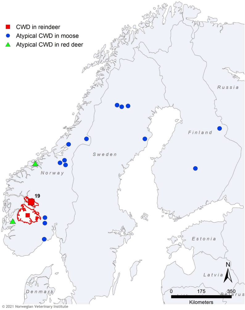
Prion diseases are fatal neurodegenerative disorders with known natural occurrence in humans and a few other mammalian species. The diseases are experimentally transmissible, and the agent is derived from the host-encoded cellular prion protein (PrPC), which is misfolded into a pathogenic conformer, designated PrPSc (scrapie). Aggregates of PrPSc molecules, constitute proteinaceous infectious particles, known as prions. Classical scrapie in sheep and goats and chronic wasting disease (CWD) in cervids are known to be infectious under natural conditions. In CWD, infected animals can shed prions via bodily excretions, allowing direct host-to-host transmission or indirectly via prion-contaminated environments. The robustness of prions means that transmission via the latter route can be highly successful and has meant that limiting the spread of CWD has proven difficult. In 2016, CWD was diagnosed for the first time in Europe, in reindeer (Rangifer tarandus) and European moose (Alces alces). Both were diagnosed in Norway, and, subsequently, more cases were detected in a semi-isolated wild reindeer population in the Nordfjella area, in which the first case was identified. This population was culled, and all reindeer (approximately 2400) were tested for CWD; 18 positive animals, in addition to the first diagnosed case, were found. After two years and around 25,900 negative tests from reindeer (about 6500 from wild and 19,400 from semi-domesticated) in Norway, a new case was diagnosed in a wild reindeer buck on Hardangervidda, south of the Nordfjella area, in 2020. Further cases of CWD were also identified in moose, with a total of eight in Norway, four in Sweden, and two cases in Finland. The mean age of these cases is 14.7 years, and the pathological features are different from North American CWD and from the Norwegian reindeer cases, resembling atypical prion diseases such as Nor98/atypical scrapie and H- and L-forms of BSE. In this review, these moose cases are referred to as atypical CWD. In addition, two cases were diagnosed in red deer (Cervus elaphus) in Norway. The emergence of CWD in Europe is a threat to European cervid populations, and, potentially, a food-safety challenge, calling for a swift, evidence-based response. Here, we review data on surveillance, epidemiology, and disease characteristics, including prion strain features of the newly identified European CWD agents.

Keywords: CWD; Deer; Fennoscandia; Moose; Nordic countries; Prion disease; Red deer; Reindeer.


Conflict of interest statement

The authors declare that they have no competing interests.

Figures

Fig. 1
Fig. 1
Distribution of CWD in reindeer, red deer, and moose in Fennoscandia as per April 2021. The outbreak of CWD among reindeer in Nordfjella, with a total of 19 cases, is indicated with a red square. The subsequent case diagnosed in a reindeer in 2020 is indicated with a smaller red square. The Nordfjella and Hardangervidda reindeer areas are marked with a red line. Atypical cases of CWD in moose are shown with blue circles and the two cases in red deer with green triangles
Fig. 2
Fig. 2
Images from the cull of the reindeer population in Nordfjella. Shot animals were transported with snowmobiles or helicopter to sites for sampling. All carcasses were destroyed with incineration after sampling
Fig. 3
Fig. 3
Immunohistochemical labelling of PrPSc in the brain of reindeer and moose diagnosed with CWD. a, b Reindeer, medulla oblongata, strong, extracellular thin and coarse granular, coalescing and plaque-like PrPSc deposition (SAF84 mAb). c, d Atypical CWD in a Norwegian moose, medulla oblongata, predominance of intraneuronal PrPSc deposition (L42 mAb). e, f Atypical CWD in a Swedish moose, thalamus, intraneuronal PrPSc deposition (SAF84 mAb)
Fig. 4
Fig. 4
Western blots analysis and interpretation of protease K-treated PrPSc (PrPres) from Norwegian reindeer and moose diagnosed with CWD, and from a bovine with classical BSE (BSE-C). a Replica blots analysed with mAbs recognizing different epitopes on PrP, as indicated below each blot. The epitopes of the mAbs are shown in c The positions of molecular weight (MW) markers are indicated on the right of the blots (in kilodaltons). Reindeer CWD is detected by the N-terminal mAb 12B2, while moose CWD and BSE-C are not. With mAb 9A2 all samples show a three-banding pattern representing di-, mono- and un-glycosylated PrPres, whose molecular weight is lower in moose than in reindeer (“Main fragment” in b and c). In contrast, mAbs directed to more C-terminal epitopes, L42 and SAF84, recognize additional low molecular weight PrPres fragments at ~ 13 and 16 kDa, detected in moose (referred to as CTF16 and CTF13 in b and c), which are glycosylated too (51). b Cartoons representing PrPres fragments detected in moose with CWD. Main, CTF16, CTF13 fragments are represented separately, with their di-glycosylayed (D), mono-glycosylated (M) and un-glycosylated (U) bands. On the right, the cartoons representing the different PrPres fragments detected in moose PrPSc are joined to show the interpretation of the complex WB patterns observed with SAF84 and L42. The position of the cartoons is in accord with the position of the bands in the blots shown in a. c. Cartoons representing full-length PrP and the different PrPres fragments detected in moose, with the position of the epitopes recognised by the mAbs used in a (ovine PrP numbering: SAF84, aa 167–173; L42, aa 148–153; 9A2, aa 99–101; 12B2, aa 93–97). Note that the 12B2 epitope is shaded in the main PrPres fragment to indicate the variability in the N-terminal Proteinase K-cleavage observed in moose with CWD. Yellow circles labelled with “N” represent the position of N-linked sugars. The GPI anchor is represented at the C-terminus

References

    1. Prusiner SB, Scott MR, DeArmond SJ, Cohen FE. Prion protein biology. Cell. 1998;93:337–348. - PubMed
    1. Silveira JR, Raymond GJ, Hughson AG, Race RE, Sim VL, Hayes SF, et al. The most infectious prion protein particles. Nature. 2005;437:257–261. - PMC - PubMed
    1. Prusiner SB. Prions. Proc Natl Acad Sci U S A. 1998;95:13363–13383. - PMC - PubMed
    1. Brandner S, Raeber A, Sailer A, Blattler T, Fischer M, Weissmann C, et al. Normal host prion protein (PrPC) is required for scrapie spread within the central nervous system. Proc Natl Acad Sci U S A. 1996;93:13148–13151. - PMC - PubMed
    1. Bueler H, Aguzzi A, Sailer A, Greiner RA, Autenried P, Aguet M, et al. Mice devoid of PrP are resistant to scrapie. Cell. 1993;73:1339–1347. - PubMed