. 2021 Nov 25;12(1):589.
doi: 10.1186/s13287-021-02656-4.

PINK1 deficiency impairs osteoblast differentiation through aberrant mitochondrial homeostasis

Affiliations

PINK1 deficiency impairs osteoblast differentiation through aberrant mitochondrial homeostasis

So-Young Lee et al. Stem Cell Res Ther. .

Abstract

Background: PTEN-induced kinase 1 (PINK1) is a serine/threonine-protein kinase in mitochondria that is critical for mitochondrial quality control. PINK1 triggers mitophagy, a selective autophagy of mitochondria, and is involved in mitochondrial regeneration. Although increments of mitochondrial biogenesis and activity are known to be crucial during differentiation, data regarding the specific role of PINK1 in osteogenic maturation and bone remodeling are limited.

Methods: We adopted an ovariectomy model in female wildtype and Pink1-/- mice. Ovariectomized mice were analyzed using micro-CT, H&E staining, Masson's trichrome staining. RT-PCR, western blot, immunofluorescence, alkaline phosphatase, and alizarin red staining were performed to assess the expression of PINK1 and osteogenic markers in silencing of PINK1 MC3T3-E1 cells. Clinical relevance of PINK1 expression levels was determined via qRT-PCR analysis in normal and osteoporosis patients.

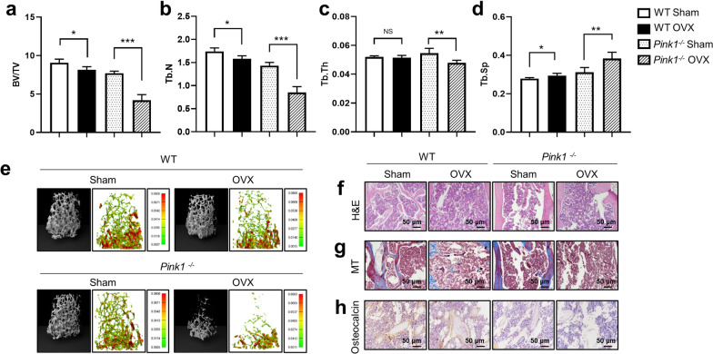
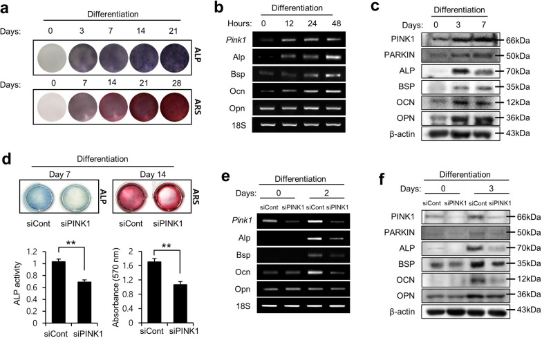
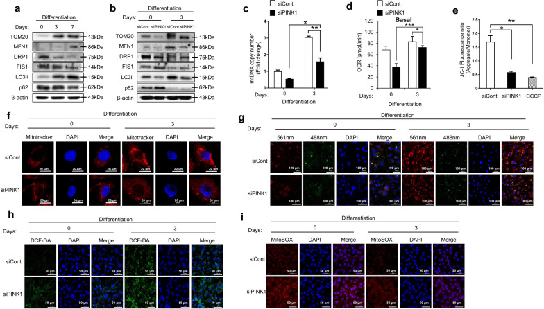
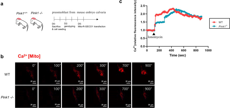
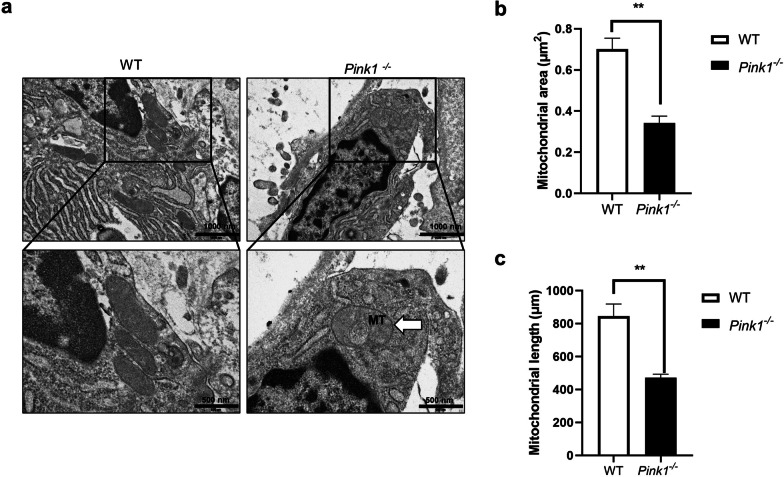
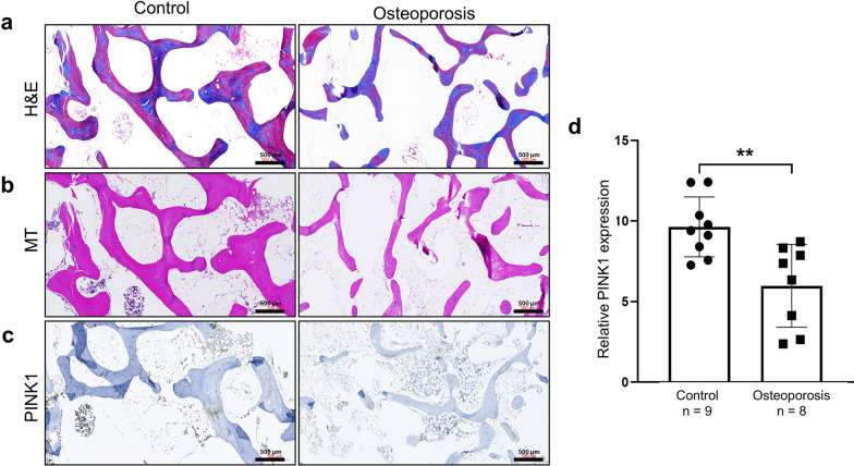
Results: A significant decrease in bone mass and collagen deposition was observed in the femurs of Pink1-/- mice after ovariectomy. Ex vivo, differentiation of osteoblasts was inhibited upon Pink1 downregulation, accompanied by impaired mitochondrial homeostasis, increased mitochondrial reactive oxygen species production, and defects in mitochondrial calcium handling. Furthermore, PINK1 expression was reduced in bones from patients with osteoporosis, which supports the practical role of PINK1 in human bone disease.

Conclusions: In this study, we demonstrated that activation of PINK1 is a requisite in osteoblasts during differentiation, which is related to mitochondrial quality control and low reactive oxygen species production. Enhancing PINK1 activity might be a possible treatment target in bone diseases as it can promote a healthy pool of functional mitochondria in osteoblasts.

Keywords: Mitochondria; Osteogenesis; Osteoporosis; PINK1.


Conflict of interest statement

The authors declare that they have no competing interests.

Figures

Fig. 1
Fig. 1
Increased bone loss in Pink1−/− mice after ovariectomy (OVX). The following parameters were calculated during the analysis of trabecular bone and architecture: a bone volume per total volume (BV/TV), b trabecular number (Tb.N), c trabecular thickness (Tb.Th), and d trabecular spacing (Tb.Sp). e Representative images of μCT and finite element analysis of femoral trabecular bones. Representative photographs of f hematoxylin–eosin (H&E), g Masson’s trichrome (MT), and h immunohistochemical staining of osteocalcin in femurs from WT or Pink1–/– mice after OVX or sham operation. Data have been expressed as mean ± SEM; *P < 0.05; **P < 0.01; ***P < 0.001; n = 6; scale bar = 50 μm
Fig. 2
Fig. 2
PINK1 is required for osteoblast differentiation. a MC3T3-E1 cells were induced to differentiate, followed by staining of the cells using alkaline phosphate (Alp) and Alizarin Red S (ARS). b mRNA levels of Pink1 and other markers of mature osteoblasts in MC3T3-E1 cells after osteogenic induction. c Protein levels of PINK1 and other osteogenic markers were analyzed using western blotting after osteogenic induction. d Alp and ARS staining of MC3T3-E1 cells that were differentiated after treatment with Pink1 siRNA. Histograms show ALP activity and quantification of Alizarin red S staining by spectrophotometry. e The effects of Pink1 siRNA on the mRNA levels of marker genes of mature osteoblasts. f The effects of Pink1 siRNA on protein levels of osteogenic markers in cells. Bsp, bone sialoprotein; Ocn, osteocalcin; Opn, osteopontin; Pink1, PTEN-induced kinase 1
Fig. 3
Fig. 3
Effect of Pink1 siRNA on mitochondrial homeostasis and ROS production during osteoblast differentiation. a Expression levels of mitochondria functional unit (Tom20), profusion (Mfn1), pro-fission (Drp1 and Fis1), and autophagy (LC3ii and p62) related proteins in MC3T3-E1 cells after osteogenic induction. b The effects of Pink1 siRNA on protein levels of mitochondria functional unit, profusion, pro-fission, and autophagy-related proteins in cells after differentiation for 3 days. c mtDNA copy number in cells after treatment with Pink1 siRNA. d The effects of Pink1 siRNA on Mitochondrial respiration, reflected by the oxygen consumption rate (OCR) level in cells after differentiation for 3 days. The oxygen consumption rate (OCR) was analyzed using a Seahorse XF-24 analyzer. Rates of basal respiration were quantified by normalization of OCR levels to total protein levels obtained from O.D. values. e Mitochondrial membrane potential (ΔΨm) was studied by measuring JC-1 uptake in MC3T3-E1 cells after osteogenic induction with or without Pink1 siRNA treatment. f Representative confocal images of MitoTracker™ (red) in MC3T3-E1 cells with or without treatment with Pink1 siRNA. Scale bar = 20 μm. g Representative confocal images of mtKeima in MC3T3-E1 cells with or without treatment with Pink1 siRNA. Scale bar = 20 μm. hi) Representative confocal images of 2,7-dichlorodihydrofluorescein diacetate (H2-DCFDA; green) and MitoSOX™ (red), showing mitochondrial and intracellular ROS production in MC3T3-E1 cells with or without treatment with Pink1 siRNA. mtDNA, mitochondrial DNA; CCCP, carbonyl cyanide m-chlorophenyl hydrazone. Data have been expressed as mean ± SEM; *P < 0.05; **P < 0.01; n = 4; scale bar = 50 μm
Fig. 4
Fig. 4
PINK1 deficiency impairs mitochondrial calcium uptake. a Experimental protocol for live cell imaging of preosteoblasts from mouse embryo calvaria. b Fluorescent time-lapse images of mito-LAR-GECO1.2-expressing preosteoblasts at the indicated times post-ionomycin treatment (500 nM). c Quantified intensity of Mito-R-GECO1 fluorescence in each indicated preosteoblast. Data from three representative cells have been shown for each treatment. Trajectories for each cell have been represented in a different color. Fluorescence intensity, quantified as average pixel intensity per cell, using the same confocal imaging system. Scale bar = 20 μm
Fig. 5
Fig. 5
Morphology of mitochondria in osteoblasts and osteocytes of femurs from Pink1−/− and WT mice. a Representative TEM of femurs from Pink1−/− and WT mice. Boxed regions have been shown in an enlarged version. Mitophagy has been indicated using arrows. Upper image scale bar = 1000 nm, lower image scale bar = 500 nm (bc). Quantitative analyses of morphometric data from TEM images have been depicted. TEM, Transmission electron microscope; MT, mitophagy. Data have been expressed as mean ± SEM; **P < 0.01; n = 3 per condition
Fig. 6
Fig. 6
Downregulation of PINK1 expression in bones of patients with osteoporosis. Representative photographs of a hematoxylin–eosin (H&E), b Masson’s trichrome (MT) and c Immunohistochemical of PINK1 staining in femurs from patients with or without osteoporosis. d Comparison of PINK1 expression in femurs from patients with or without osteoporosis. Data have been expressed as mean ± SEM; **P < 0.01; scale bar = 500 μm

References

    1. Boyle WJ, Simonet WS, Lacey DL. Osteoclast differentiation and activation. Nature. 2003;423:337–342. doi: 10.1038/nature01658. - DOI - PubMed
    1. Ikeda K, Takeshita S. Factors and mechanisms involved in the coupling from bone resorption to formation: how osteoclasts talk to osteoblasts. J Bone Metab. 2014;21:163–167. doi: 10.11005/jbm.2014.21.3.163. - DOI - PMC - PubMed
    1. Hou Z, et al. KLF2 regulates osteoblast differentiation by targeting of Runx2. Lab Invest. 2019;99:271–280. doi: 10.1038/s41374-018-0149-x. - DOI - PubMed
    1. Lee SY, et al. PGC1alpha activators mitigate diabetic tubulopathy by improving mitochondrial dynamics and quality control. J Diabetes Res. 2017;2017:6483572. doi: 10.1155/2017/6483572. - DOI - PMC - PubMed
    1. Kain KH, Popov VL, Herzog NK. Alterations in mitochondria and mtTFA in response to LPS-induced differentiation of B-cells. Biochim Biophys Acta. 2000;1494:91–103. doi: 10.1016/s0167-4781(00)00223-2. - DOI - PubMed

Publication types

Substances