. 2021 Nov 25;11(1):22955.
doi: 10.1038/s41598-021-02448-z.

Bone defect map of the true acetabulum in hip dysplasia (Crowe type II and III) based on three-dimensional image reconstruction analysis

Affiliations

Bone defect map of the true acetabulum in hip dysplasia (Crowe type II and III) based on three-dimensional image reconstruction analysis

Xinggui Wen et al. Sci Rep. .

Abstract

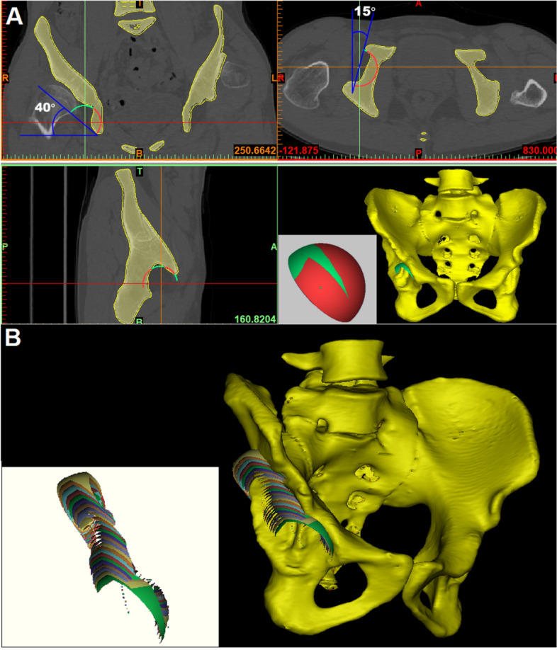
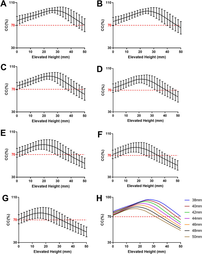
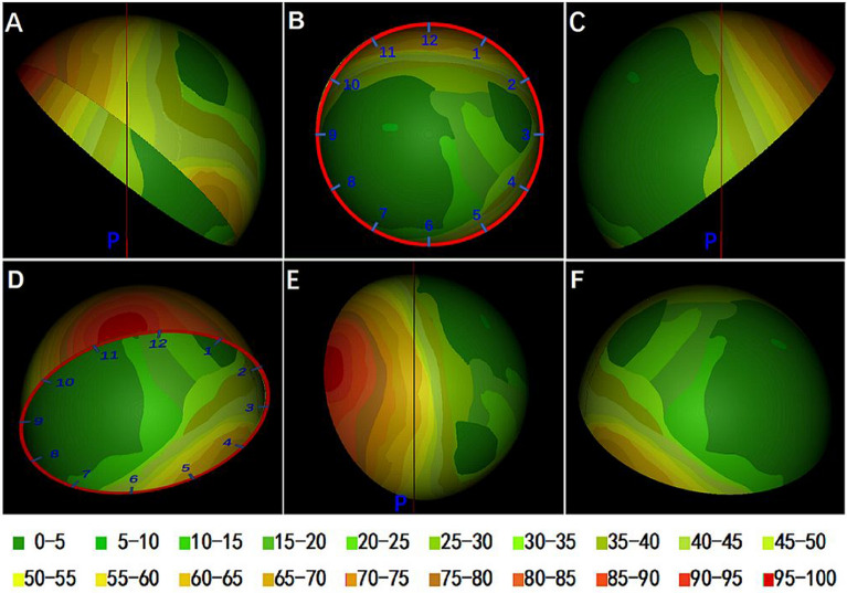
The high hip center technique (HHC) is considered to be feasible for acetabular reconstruction in patients with DDH, but there is little in-depth study of its specific impact on Crowe type II and III DDH. The purpose of this study was to simultaneously analyze the effect of HHC on bone coverage of the cup (CC) in the acetabular reconstruction of type II and III DDH patients and to propose a map of acetabular bone defects from the perspective of the cup. Forty-nine hip CT data of 39 patients with DDH (Crowe type II and III) were collected to simulate acetabular reconstruction by cup models of different sizes (diameter 38mm-50 mm, 2 mm increment) with the HHC technique. The frequency distribution was plotted by overlapping the portions of the 44 mm cups that were not covered by the host bone. The mean CC of cups with sizes of 38 mm, 40 mm, 42 mm, 44 mm, 46 mm, 48 mm, and 50 mm at the true acetabula were 77.85%, 76.71%, 75.73%, 74.56%, 73.68%, 72.51%, and 71.75%, respectively, and the maximum CC increments were 21.24%, 21.58%, 20.86%, 20.04%, 18.62%, 17.18%, and 15.42% (P < 0.001), respectively, after the cups were elevated from the true acetabula. The bone defect map shows that 95% of type II and III DDH acetabula had posterosuperior bone defects, and approximately 60% were located outside the force line of the hip joint. Acetabular cups can meet a CC of more than 70% at the true acetabulum, and approximately 60% of Crowe type II and III DDH patients can obtain satisfactory CC at the true acetabulum by using a 44-mm cup without additional operations.


Conflict of interest statement

The authors declare no competing interests.

Figures

Figure 1
Figure 1
3D images of the pelvis reconstructed by Mimics software (Materialise, Version 17.0.0.435, RUL: https://www.materialise.com/en/medical/mimics-innovation-suite/mimics) using complete CT data of the pelvis. (A) The cup models were placed in the 3D reconstruction image of the pelvis to simulate acetabular reconstruction in THA. The position of the cup was adjusted to 40° of abduction and 15° of anteversion on the coronal and transverse sections. The total surface area of the cup (St) can be read directly from the Mimics software. Boolean operation was used to obtain the uncovered area of the cup (Su), which is indicated in green; the covered area is indicated in red. (B) The uncovered portions of the cup with a diameter of 50 mm at different elevations are indicated in different colors.
Figure 2
Figure 2
Changes in CC and the elevated height of the cup from the true acetabulum for cup sizes of 38 mm (A), 40 mm (B), 42 mm (C), 44 mm (D), 46 mm (E), 48 mm (F), and 50 mm (G). H shows the relationship between the CC and the elevated height of cups of different sizes. With increasing cup size, the elevated height of the cup decreased gradually when the maximum CC was reached.
Figure 3
Figure 3
Relationship of the CC and elevated height of cups from the true acetabulum until the CC reached the maximum value. PC: Pearson correlation coefficient.
Figure 4
Figure 4
The distribution frequency of the uncovered portion on intact cup is shown in different colors. The result is expressed as a percentage with a total of 49. A clock pattern was drawn on the cup to show the location of the uncovered portion. A plane perpendicular to the anterior plane of the pelvis was built through the rotation center of the cup. (A) Front view of the pelvis. The red line is a vertical line through the rotation center of the mortar cup. (B) Front view of the cup. (C) Rear view of the cup. (D) Vertical view of the cup. p: Vertical plane through the rotation center of the cup.

References

    1. Crowe JF, Mani VJ, Ranawat CS. Total hip replacement in congenital dislocation and dysplasia of the hip. J. Bone Joint Surg. Am. 1979;61(1):15–23. doi: 10.2106/00004623-197961010-00004. - DOI - PubMed
    1. Greber EM, Pelt CE, Gililland JM, Anderson MB, Erickson JA, Peters CL. Challenges in total hip arthroplasty in the setting of developmental dysplasia of the hip. J. Arthroplasty. 2017;32(9s):S38–s44. doi: 10.1016/j.arth.2017.02.024. - DOI - PubMed
    1. Chougle A, Hemmady MV, Hodgkinson JP. Severity of hip dysplasia and loosening of the socket in cemented total hip replacement. J. Bone Joint Surg. Br. 2005;87(1):16–20. doi: 10.1302/0301-620X.87B1.15199. - DOI - PubMed
    1. Hartofilakidis G, Karachalios T. Total hip arthroplasty for congenital hip disease. J. Bone Joint Surg. Am. 2004;86(2):242–250. doi: 10.2106/00004623-200402000-00005. - DOI - PubMed
    1. Hartofilakidis G, Stamos K, Karachalios T. Treatment of high dislocation of the hip in adults with total hip arthroplasty: Operative technique and long-term clinical results. J. Bone Joint Surg. Am. 1998;80(4):510. doi: 10.2106/00004623-199804000-00007. - DOI - PubMed

Publication types

MeSH terms