Review
. 2022 Jan;23(1):21-39.
doi: 10.1038/s41580-021-00432-z. Epub 2021 Nov 25.

Structures and functions of coronavirus replication-transcription complexes and their relevance for SARS-CoV-2 drug design

Affiliations
Review

Structures and functions of coronavirus replication-transcription complexes and their relevance for SARS-CoV-2 drug design

Brandon Malone et al. Nat Rev Mol Cell Biol. 2022 Jan.

Abstract

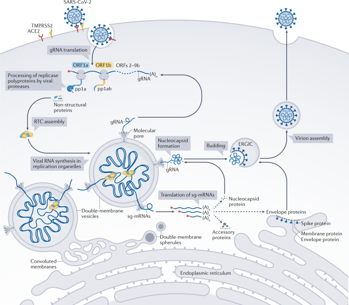
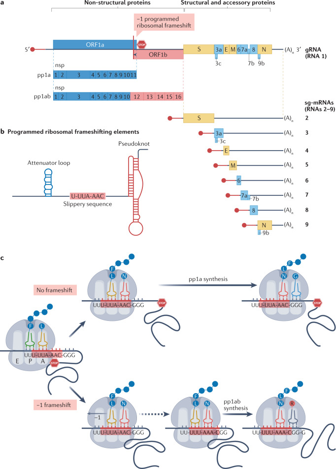
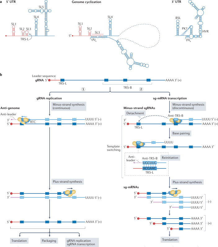
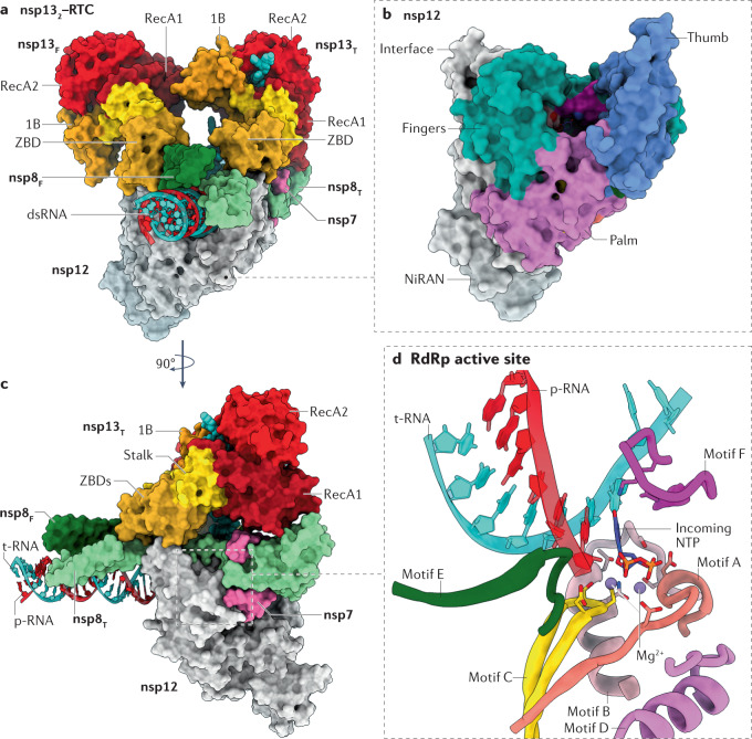
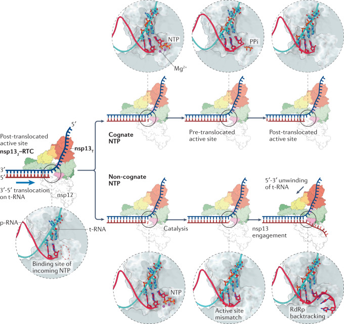
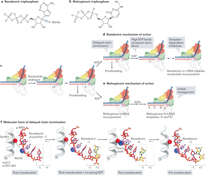
Severe acute respiratory syndrome coronavirus 2 (SARS-CoV-2) has killed millions of people and continues to cause massive global upheaval. Coronaviruses are positive-strand RNA viruses with an unusually large genome of ~30 kb. They express an RNA-dependent RNA polymerase and a cohort of other replication enzymes and supporting factors to transcribe and replicate their genomes. The proteins performing these essential processes are prime antiviral drug targets, but drug discovery is hindered by our incomplete understanding of coronavirus RNA synthesis and processing. In infected cells, the RNA-dependent RNA polymerase must coordinate with other viral and host factors to produce both viral mRNAs and new genomes. Recent research aiming to decipher and contextualize the structures, functions and interplay of the subunits of the SARS-CoV-2 replication and transcription complex proteins has burgeoned. In this Review, we discuss recent advancements in our understanding of the molecular basis and complexity of the coronavirus RNA-synthesizing machinery. Specifically, we outline the mechanisms and regulation of RNA translation, replication and transcription. We also discuss the composition of the replication and transcription complexes and their suitability as targets for antiviral therapy.


Conflict of interest statement

E.A.C has received funding from Gilead Sciences to fund research on remdesivir’s incorporation into the RNA-dependent RNA polymerase. The other authors declare no competing interests.

Figures

Fig. 1
Fig. 1. The SARS-CoV-2 infection cycle.
To enter a host cell, the severe acute respiratory syndrome coronavirus 2 (SARS-CoV-2) spike protein interacts with the cellular surface protein angiotensin-converting enzyme 2 (ACE2), while being cleaved by cellular proteases such as transmembrane serine protease 2 (TMPRSS2) to activate its membrane-fusion capacity. The genomic RNA (gRNA), which is capped on its 5′ end (red circle) and polyadenylated ((A)n) on its 3′ end, is released from the viral particle and — after recruiting host-cell ribosomes — translated into two replicase polyproteins, pp1a and pp1ab. Proteases embedded in viral non-structural protein 3 (nsp3) and nsp5 cleave pp1a and pp1ab into 16 non-structural proteins that assemble into replication–transcription complexes (RTCs). Viral RNA synthesis occurs within double-membrane vesicles that are part of virus-induced membranous replication organelles (Box 1). The RTCs produce new gRNAs and a set of subgenomic mRNAs (sg-mRNAs) that include open reading frames (ORFs) 2–9b, which encode the structural spike, membrane, envelope and nucleocapsid proteins, and also a number of accessory proteins. Newly made gRNAs can be translated to yield additional non-structural proteins, serve as a template for further RNA synthesis or be packaged into new virions. SARS-CoV-2 assembly starts with the coating of gRNAs with nucleocapsid proteins, generating nucleocapsid structures that bud into the endoplasmic reticulum–Golgi intermediate compartment (ERGIC), thereby acquiring a lipid bilayer containing the viral spike, membrane and envelope proteins. Adapted from ref., CC BY 4.0 (https://creativecommons.org/licenses/by/4.0/).
Fig. 2
Fig. 2. Regulation of SARS-CoV-2 gene expression on the translational level.
a | Severe acute respiratory syndrome coronavirus 2 (SARS-CoV-2) genome organization. Open reading frames (ORFs) are not drawn exactly to scale. The SARS-CoV-2 genomic RNA (gRNA) has a 5′ cap (red circle) followed by a leader sequence (red line) that is shared with all subgenomic mRNAs (sg-mRNAs), and a 3′ poly(A) tail. The 5′-proximal three quarters of the genome encode the replicase polyproteins pp1a and pp1ab, which are cleaved to yield 16 non-structural proteins (nsp1–nsp16; blue from ORF1a and red from ORF1b). The 3′-proximal one quarter of the genome encodes the structural (brown) and accessory (azure) proteins. Structural and accessory proteins are expressed from a nested set of sg-mRNAs, with ORF3c, ORF7b and ORF9b being expressed via ribosomal ‘leaky scanning’. b | RNA motif and structures that promote a frameshift from ORF1a to ORF1b, thereby controlling the synthesis of pp1ab. The key programmed ribosomal frameshifting (PRF)-stimulating RNA structure — a pseudoknot — interacts with the ribosome and induces its pausing, which generates tension in the gRNA template. As a result, ribosomes can slip one nucleotide backwards on the ‘slippery sequence’ (−1 PRF). An attenuating RNA loop located upstream of the slippery sequence also contributes to modulating PRF frequency. c | Model of −1 PRF at the ORF1a–ORF1b junction, showing the regulatory RNA elements inducing a simultaneous −1 shift of the tRNAs bound to the A and P sites of the ribosome, which can then translate ORF1b. The one-letter code for amino acids (circles) is used. A stop sign represents the ORF1a stop codon. E, envelope protein; M, membrane protein; N, nucleocapsid protein; S, spike protein.
Fig. 3
Fig. 3. SARS-CoV-2 RNA replication and transcription.
a | The genomic RNA (gRNA) of severe acute respiratory syndrome coronavirus 2 (SARS-CoV-2) has cis-acting structures at its 5′ untranslated region (UTR) and 3′ UTR, and engages in long-distance intramolecular RNA–RNA interactions; these structures and interactions are thought to be involved in the regulation of replication and transcription. The 5′ UTR contains five conserved stem–loop (SL) structures, and the 3′ UTR contains a bulged stem–loop (BSL), a (predicted) pseudoknot (PK) and a stem–loop structure with a hypervariable region (HVR). SARS-CoV-2 gRNA cyclization results in complete opening of SL3, where the leader transcription regulatory sequence (TRS-L) resides. b | The gRNA serves as a template for gRNA replication (step 1) and for subgenomic mRNA (sg-mRNA) transcription (step 2); each process requires dedicated minus-strand templates: the anti-genome and a set of minus-strand sgRNAs, respectively. Synthesis of the latter involves a discontinuous step in which the replication–transcription complex (RTC) pauses RNA synthesis after copying one of the body transcription regulatory sequences (TRS-B), and detaches from the template. Subsequently, the RTC relocates to a position near the 5′ end of the gRNA template, where the complement of the TRS-B (anti-TRS-B) in the nascent minus-strand sgRNA engages in base pairing with the TRS-L. This template switch leads to the addition of the complement of the gRNA leader sequence (anti-leader) to the 3′ end of each of minus-strand sgRNA, which are used as templates for sg-mRNA production, thereby ensuring that all coronavirus sg-mRNAs include a 5′-terminal leader sequence of ~75 nucleotides that is identical to the 5′-terminal sequence of the gRNA. Positions of the TRSs and anti-TRSs are schematic and not drawn exactly to scale. Part b adapted from ref., CC BY 4.0 (https://creativecommons.org/licenses/by/4.0/).
Fig. 4
Fig. 4. Architecture of SARS-CoV-2 RNA replication and transcription complexes.
a | Surface-rendered representation illustrating two molecules of non-structural protein 13 (nsp13) bound to the replication–transcription complex (nsp132–RTC) of severe acute respiratory syndrome coronavirus 2 (SARS-CoV-2). Depicted are the nsp7–nsp8–nsp12 RNA-dependent RNA polymerase (RdRp) holoenzyme bound to a double-stranded RNA (dsRNA) scaffold and to two molecules of nsp13: nsp13 thumb (nsp13T) and nsp13 fingers (nsp13F). The 1B, RecA1 and RecA2 domains of nsp13 are highlighted. b | The nsp12 RdRp domain adopts the canonical conformation of a ‘cupped right hand’ and its structural motifs are named accordingly. The nidovirus RdRp-associated nucleotidyltransferase (NiRAN) domain constitutes the amino-terminal 250 residues of nsp12. c | View of the nsp132–RTC complex rotated by 90° compared with part a. d | Zoomed-in view of the RdRp active site and its conserved structural motifs, except for motif G, which was excluded from the illustration for clarity. The active site is depicted with an incoming nucleotide, modelled using the pre-incorporation structures of the hepatitis C virus RdRp (Protein Data Bank entry 4WTL). Residues involved in both chelating the catalytic Mg2+ ions and orientating the incoming nucleotide are shown in a stick representation. Protein Data Bank entry 6XEZ was used in preparation of this figure. NTP, nucleoside triphosphate; p-RNA, product RNA; t-RNA, template RNA; ZBD, zinc-binding domain.
Fig. 5
Fig. 5. The nucleotide addition cycle.
Model of the interactions between non-structural protein 13 thumb (nsp13T) and nsp12 during RNA synthesis. Zoomed-in views of the active site during the nucleotide addition cycle are presented above or below each panel. In the presence of a natural, cognate nucleoside triphosphate (NTP), the incoming NTP forms a phosphodiester bond with the product RNA (p-RNA) (top). Nsp13T is not engaged with the template RNA (t-RNA), and forward translocation of the nsp12 RNA-dependent RNA polymerase (RdRp) on the t-RNA strand is unimpeded. In the event of incorporation of a non-natural or non-cognate nucleotide, the active site is perturbed and activates the helicase nsp13 to induce RdRp backtracking, as shown in the last panel (bottom). nsp132, nsp13T–nsp13F; PPi, pyrophosphate; RTC, replication–transcription complex.
Fig. 6
Fig. 6. Mechanisms of inhibition of coronavirus RNA synthesis by remdesivir and molnupiravir.
a | Chemical structure of remdesivir triphosphate, the activated form of remdesivir (Veklury). The ribose 1′-nitrile group is highlighted. b | Chemical structure of molnupiravir triphosphate, the activated form of molnupiravir (EIDD-2801/MK-4482). c | Schematic showing initial recognition and incorporation of a nucleotide analogue. ‘+1’ refers to the position of the nucleotide analogue before incorporation, whereas ‘−1’ refers to the position of the nucleotide analogue following catalysis and movement into the post-translocated register. d | Remdesivir exerts its inhibitory effect through ‘delayed chain termination’ and ‘template-dependent inhibition’,. Delayed chain termination impedes RNA-dependent RNA polymerase (RdRp) translocation through a steric clash that occurs when remdesivir reaches the fourth position (see part f) from the 3′ end of the product RNA (p-RNA) strand (red strand). Delayed chain termination is alleviated by high nucleoside trisphosphate (NTP) concentrations, leading to the proposal of template-dependent inhibition, which occurs when remdesivir is positioned at the RdRp active site in the template RNA (t-RNA) strand. e | Molnupiravir perturbs replication through ‘lethal mutagenesis’, by enabling the indiscriminate incorporation of either ATP (A) or GTP (G) when it is positioned in the t-RNA strand,. f | Structural analysis of the delayed chain termination mechanism. Following incorporation of remdesivir and its translocation to the −3 active site position, the ribose 1′-nitrile group is positioned to collide with the side chain of non-structural protein 12 (nsp12) Ser861 (expected clash shown by inhibitory arrows). Addition of the next nucleotide triggers the steric clash of the Ser861 side chain and the remdesivir nitrile group, forcing the growing RNA chain to populate the ‘pre-translocated’ state. High NTP concentrations may alleviate the energetic cost imposed by the translocation barrier, by occupying the +1 site and thereby driving the forward movement of the RdRp. The structural models are based on Protein Data Bank entries 7B3B, 7B3C and 7B3D.

Similar articles

Cited by

References

    1. Coronaviridae Study Group of the International Committee on Taxonomy of Viruses. The species Severe acute respiratory syndrome-related coronavirus: classifying 2019-nCoV and naming it SARS-CoV-2. Nat. Microbiol. 2020;5:536–544. doi: 10.1038/s41564-020-0695-z. - DOI - PMC - PubMed
    1. Gulyaeva AA, Gorbalenya AE. A nidovirus perspective on SARS-CoV-2. Biochem. Biophys. Res. Commun. 2021;538:24–34. doi: 10.1016/j.bbrc.2020.11.015. - DOI - PMC - PubMed
    1. Gorbalenya AE, Enjuanes L, Ziebuhr J, Snijder EJ. Nidovirales: evolving the largest RNA virus genome. Virus Res. 2006;117:17–37. doi: 10.1016/j.virusres.2006.01.017. - DOI - PMC - PubMed
    1. Snijder, E. J., Decroly, E. & Ziebuhr, J. in Advances in Virus Research Vol. 96 (ed. Ziebuhr, J.) 59–126 (Elsevier, 2016). - PMC - PubMed
    1. Saberi A, Gulyaeva AA, Brubacher JL, Newmark PA, Gorbalenya AE. A planarian nidovirus expands the limits of RNA genome size. PLOS Pathog. 2018;14:e1007314. doi: 10.1371/journal.ppat.1007314. - DOI - PMC - PubMed

Publication types