Clinical Trial
. 2021 Nov 19:13:479-494.
doi: 10.2147/CCIDE.S332687. eCollection 2021.

Radiographic and Esthetic Evaluation Following Immediate Implant Placement with or without Socket Shield and Delayed Implant Placement Following Socket Preservation in the Maxillary Esthetic Region - A Randomized Controlled Clinical Trial

Affiliations
Clinical Trial

Radiographic and Esthetic Evaluation Following Immediate Implant Placement with or without Socket Shield and Delayed Implant Placement Following Socket Preservation in the Maxillary Esthetic Region - A Randomized Controlled Clinical Trial

Muthukumar Santhanakrishnan et al. Clin Cosmet Investig Dent. .

Abstract

Objective: The purpose of this study was assessment of the changes in soft and hard tissues in the esthetic zone of maxilla following immediate implant placement (IIP) with and without the socket shield technique (SST) and placement of implants 4 months following socket preservation (DIP) in terms of alterations in crestal bone thickness (CBT) and soft tissue changes evaluated by means of pink esthetic scores (PES) following placement of implants in the esthetic zone of maxilla.

Materials and methods: In the maxillary esthetic region, 75 dental implants were placed totally, with 25 implants each in the SST, IIP, and DIP groups. All participants were subjected to undergo CBCT for assessing the variations in thickness of crestal aspect of facial/buccal/labial alveolar bone (CBT). PES and PROMS (patient-related outcome measures) were assessed using VAS for pain threshold and esthetic satisfaction following implant placement and after 6th post-operative month.

Results: The mean reduction in CBT showed a statistically significant difference between and within the groups, in comparison to IIP and DIP groups, which demonstrated an average reduction in CBT 0.4 ± 0.1 and 0.2 ± 0.1 at 6 months following implant placement, respectively. The SST group showed a significantly lesser reduction in CBT of 0.05 ± 0.02. However, the mean difference in PES within and among the groups showed no significant difference statistically at P < 0.05. On comparison of individual scores of PES between the groups, the results showed significant difference statistically at P < 0.001.

Conclusion: The SST group demonstrated minimal reduction in CBT and a superior PES at the end of 6 months compared with the IIP and DIP groups.

Keywords: immediate implant placement; pink esthetic score; randomized controlled trial; socket preservation; socket shield technique.


Conflict of interest statement

The authors report no conflicts of interest in this work.

Figures

Figure 1
Figure 1
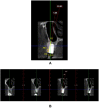
Original. Demonstration of a treated case, Radiographic measurements. (A) Pre-operative CBCT - sagittal view. (B) Thickness of labial plate assessed in section of 1 mm.
Figure 2
Figure 2
Original. CONSORT flow chart (n) representing the implant sites.
Figure 3
Figure 3
Original. Socket shield technique and implant placement in relation to tooth no 14. (A) Pre-operative site in relation to tooth no 14. (B) Root stumps present in relation to tooth no 14. (C) The buccal root fragment prepared according to the SST protocol and the implant in position with the “jumping distance” grafted. (D) Post-operative IOPA (intra-oral periapical radiograph) showing implant in position. (E) Post-operative view of implant in relation to tooth no 14. (F) S-shaped emergence profile of the provisional restoration. (G) The provisional restoration placed post-operatively.
Figure 4
Figure 4
Original. Immediate implant placement in relation to tooth no 15. (A) Pre-operative site in relation to tooth no 15. (B) Gross destruction present in relation to tooth no 15. (C) Atraumatic extraction of tooth no 15. (D) Socket post-extraction in relation to tooth no 15. (E) Implant placed in relation to tooth no 15. (F) Post-operative IOPA (intra-oral periapical radiograph) showing implant in position. (G) Sutures placed in relation to tooth no 15. (H) Ceramo -metal crown placed in relation to tooth no 15.
Figure 5
Figure 5
Original. Socket preservation and delayed implant placement in relation to tooth no 22. (A) Atraumatic extraction in relation to tooth no 22. (B) Socket post-extraction in relation to tooth no 22. (C and D) Clinical measurements of socket by means of a stent. (E) Preparation of A-PRF membrane. (F) DBBM and A- PRF mixed at a ratio of 1:1. (G) Outline for procurement of connective tissue graft. (H) Connective tissue graft procured from hard palate. (I) Donor site stabilized by Ab gel with a transverse mattress suture. (J) Extraction socket filled with DBBM +A-PRF. (K) Socket sealed with connective tissue graft and flaps sutured. (L) Incision outline for placement of implant in relation to tooth no 22. (M) Placement of implant in relation to tooth no 22. (N) Cover screw placed in relation to tooth no 22. (O) Post-operative IOPA (intra-oral periapical radiograph) showing implant in position. (P) Sutures placed in relation to tooth no 22.
Figure 6
Figure 6
Original. Post-operative CBCT of a treated case. (A) Sagittal view. (B) Labial cortical thickness assessed in sections of 1-mm thickness.
Figure 7
Figure 7
Original. Difference in CBT of Group I, Group II, and Group III of the study participants (n = 75). *A P value<0.05 considered as significant by the one-way ANOVA test for between-group and within-group differences was tested by repeated measures ANOVA.
Figure 8
Figure 8
Original. Changes in PES& scores of Group I, Group II, and Group III of the study participants (n = 75). The P value was tested by Friedman test for within-group differences. The P value was tested by the Kruskal–Wallis test for between-group differences.

References

    1. Hämmerle CH, Araújo MG, Simion M; Osteology Consensus Group 2011. Evidence-based knowledge on the biology and treatment of extraction sockets. Clin Oral Implants Res. 2012;23(Suppl 5):80–82. doi:10.1111/j.1600-0501.2011.02370.x - DOI - PubMed
    1. Bramanti E, Norcia A, Cicciu M, et al. Postextraction dental Implant in the aesthetic zone, socket shield technique versus conventional protocol. J Craniofac Surg. 2018;29(4):1037–1041. doi:10.1097/SCS.0000000000004419 - DOI - PubMed
    1. De Risi V, Clementini M, Vittorini G, Mannocci A, De Sanctis M. Alveolar ridge preservation techniques: a systematic review and meta-analysis of histological and histomorphometrical data. Clin Oral Implants Res. 2015;26(1):50–68. doi:10.1111/clr.12288 - DOI - PubMed
    1. Annunziata M, Guida L, Nastri L, et al. The role of autologous platelet concentrates in alveolar socket preservation: a systematic review. J Transfus Med Hemother. 2018;45:195–203. doi:10.1159/000488061 - DOI - PMC - PubMed
    1. Buser D, Chappuis V, Belser URSC, Chen S. Implant placement post extraction in esthetic single tooth sites: when immediate, when early, when late? Periodontology. 2017;73:84–102. doi:10.1111/prd.12170 - DOI - PubMed

LinkOut - more resources