

Is Flexion Gap Rectangular in Native Indian Knees? Results of an MRI Study
- PMID: 34824712
- PMCID: PMC8586387
- DOI: 10.1007/s43465-021-00418-1
Is Flexion Gap Rectangular in Native Indian Knees? Results of an MRI Study
Abstract
Background: The purpose of this study was to evaluate the flexion-gap of the native knees in the normal population and to assess any gender-specific variations in the flexion gap of the knees.
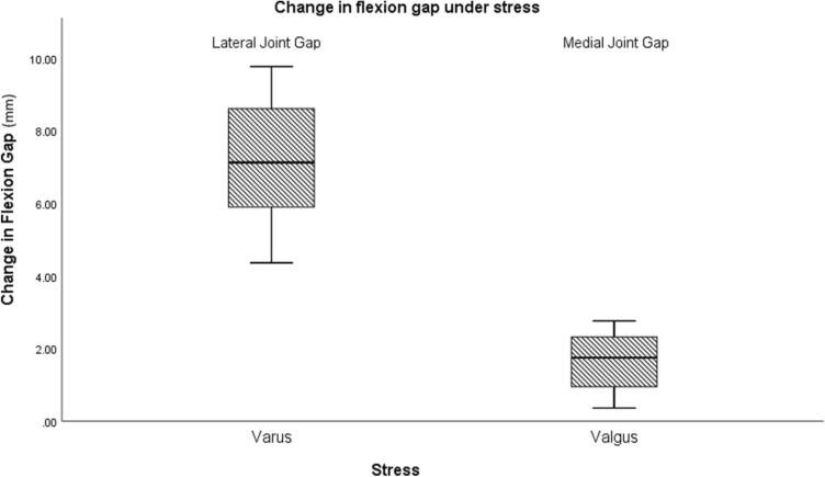
Methods: A total of 50 normal asymptomatic volunteers with normal knee radiographs were selected for MRI of the knee. The left knee was scanned in an open MRI using a T1-weighted sequence. Imaging was performed in neutral, passive varus and valgus stress at 90° of knee flexion by placing custom-made blocks on a special board consecutively below the distal part of the leg.
Results: The study population consisted of 26 males and 24 females with a mean age of 25.77 years. Under varus stress, the mean lateral flexion gap increased to 9.28 ± 1.53 mm and under valgus stress, the mean medial flexion gap increased to 2.75 ± 1.22 mm from neutral. The increase in the flexion gap on the lateral side was 5.28 ± 1.79 mm, which was significantly higher compared to that on the medial side. In gender-specific analysis, the mean lateral flexion gap was 10.21 mm in females and 8.46 mm in males under varus stress.
Conclusion: The findings of the study indicate that the lateral soft tissues are more lax compared to the medial soft tissue structures and this laxity is higher in females as compared to males. The study provides evidence of the existing physiological variations of these soft tissue structures resulting in a trapezoidal flexion gap in the native knees rather than the recommended rectangular gap.
Keywords: Knee; Laxity; MRI study; Total knee arthroplasty.
© Indian Orthopaedics Association 2021.
Conflict of interest statement
Conflict of interestThe authors have no conflicts of interest to declare.
Figures


References
-
- Willcox NMJ, Clarke JV, Smith BRK, Deakin AH, Deep K. A comparison of radiological and computer navigation measurements of lower limb coronal alignment before and after total knee replacement. The Journal of Bone and Joint Surgery Series B. 2012;94:1234–1240. doi: 10.1302/0301-620X.94B9.28250. - DOI - PubMed
-
- Scott WN. Insall & Scott surgery of the knee. 6. Churchill Livingstone/Elsevier; 2017.
-
- Scuderi G, Insall J. The posterior stabilized knee prosthesis. Orthopedic Clinics of North America. 1989;20:71–78. - PubMed
-
- Beverland D. E. (2009). Soft-tissue balance in total knee arthroplasty. In European instructional lectures. Springer Berlin Heidelberg. pp. 213–8.
LinkOut - more resources
Full Text Sources
Miscellaneous