

A Comprehensive Review of Congenital Lumbar Synostosis and Associated Findings
- PMID: 34824930
- PMCID: PMC8610779
- DOI: 10.7759/cureus.19013
A Comprehensive Review of Congenital Lumbar Synostosis and Associated Findings
Abstract
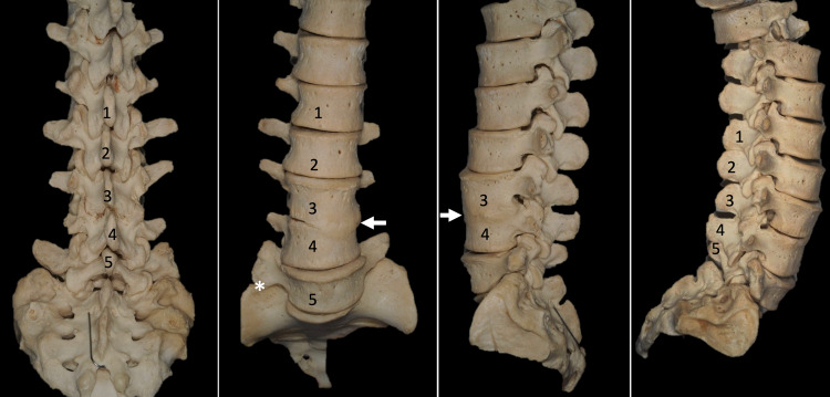
Congenital vertebral synostosis (CVS) is a rare developmental condition due to failure of vertebral segmentation. Vertebrae and their intervertebral discs differentiate and resegment at the time of organogenesis during fetal life. Failure of this embryological process can result in the limitation of mobility of the involved segment. This inappropriate segmentation thus results in vertebral fusion or a block vertebra with subsequent vertebral synostosis. Long-term, such segmental fusion can increase osteoarthritis at levels below and above the fused segment due to excessive wear on these joints. Presentations can include congenital kyphosis and scoliosis. Patients may present with back and radicular pain, and possible myelopathy CVS usually occurs, in order of frequency, in the cervical, lumbar, and thoracic vertebral levels. This paper reviews congenital lumbar synostosis with associated findings and its clinical implications and embryological significance. A case illustration is also included.
Keywords: congenital vertebral synostosis; klippel-feil syndrome; lumbar synostosis; spine; vertebral segmentation.
Copyright © 2021, Volk et al.
Conflict of interest statement
The authors have declared that no competing interests exist.
Figures

References
-
- A spectrum of vertebral synostosis. Kulkarni V, Ramesh BR. http://file:///C:/Users/703693/Downloads/12...024Kulkarni...ASpectrum...... Int J Basic Appl Med Sci. 2012;2:71–77.
-
- Adler JT. Philadelphia: Elsevier Inc; 2009. Gray's Anatomy: The Anatomical Basis of Clinical Practice, 40th Edition.
-
- Yochum TR, Rowe LJ. Baltimore: Lippincott Williams & Wilkins; 2007. Essentials of Skeletal Radiology, 3rd ed, 2 vols.
-
- The vertebral body: radiographic configurations in various congenital and acquired disorders. Kumar R, Guinto FC Jr, Madewell JE, Swischuk LE, David R. Radiographics. 1988;8:455–485. - PubMed
-
- Familial Klippel-Feil syndrome and paracentric inversion inv(8)(q22.2q23.3) Clarke RA, Singh S, McKenzie H, Kearsley JH, Yip MY. https://pubmed.ncbi.nlm.nih.gov/8533765/ Am J Hum Genet. 1995;57:1364–1370. - PMC - PubMed
Publication types
LinkOut - more resources
Full Text Sources