

Novel PLA/ZnO Nanofibrous Nanocomposite Loaded with Tranexamic Acid as an Effective Wound Dressing: In Vitro and In Vivo Assessment
- PMID: 34825013
- PMCID: PMC8590716
- DOI: 10.30498/ijb.2021.220458.2737
Novel PLA/ZnO Nanofibrous Nanocomposite Loaded with Tranexamic Acid as an Effective Wound Dressing: In Vitro and In Vivo Assessment
Abstract
Background: Chronic wounds contribute to the majority of clinical cases, associated with significant morbidity, and place a massive financial burden on healthcare systems. Thus, various bandage mats have been designed to facilitate wound healing in clinical applications. Polylactic acid (PLA) nanofibers, as suitable drug carriers, are highly desirable to prepare a controlled environment for wound healing in dressing tissue. Zinc oxide (ZnO) nanoparticles as an effective antibacterial agent for wound treatment prevent bacterial invasion and wound infection.
Objectives: In this project, for the first time, a new (PLA)/(ZnO) nanofibrous nanocomposite loaded with tranexamic acid (TXA) has been introduced as a useable dressing in wound healing. Furthermore, the antibacterial properties, coagulant assay, and wound healing assays of nanocomposite are evaluated.
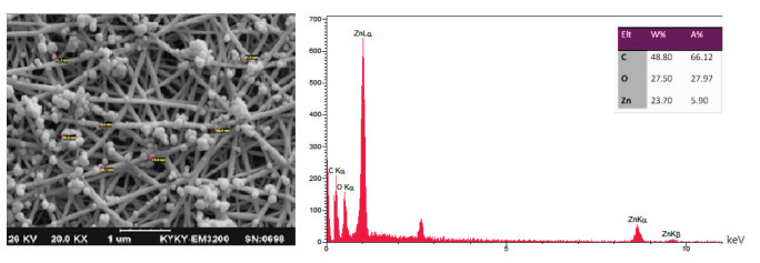
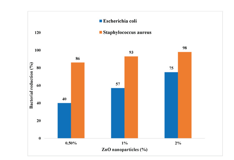
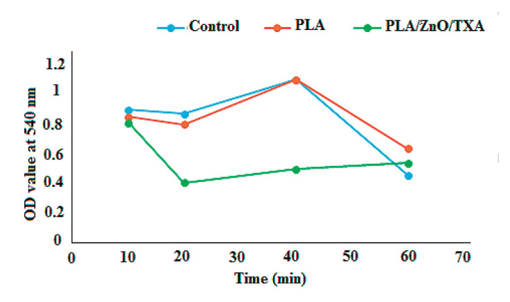
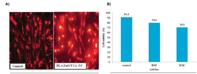
Material and methods: PLA/ZnO nanofibrous nanocomposites were loaded with tranexamic acid fabricated by electrospinning method at distinct concentrations. The prepared structure was characterized using field emission scanning electron microscopy (FESEM), energy-dispersive X-ray spectroscopy (EDS), and Fourier transform infrared spectroscopy (FTIR). Further, antimicrobial properties of tissue were investigated against Escherichia coli and Staphylococcus aureus bacteria. Also, the coagulation assays, in vitro cytotoxicity, and in vivo skin wound healing model in mice were evaluated.
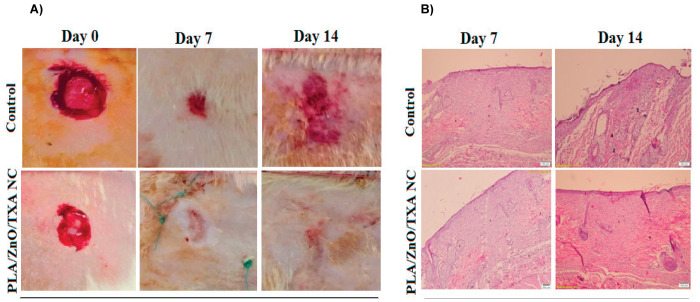
Results: Morphological analysis of the prepared nanofibrous nanocomposites showed uniform bead-free nanofibers with an average size of 90 nm diameter. The structure exhibited proper antibacterial activities against Escherichia coli and Staphylococcus aureus bacteria, and a good blood clotting effect. In vitro cytotoxicity assay of the structure approved that this mat has no cytotoxic effect on human dermal fibroblast cells. In vivo wound healing examination in mice observed over 7 and 14 days showed a faster rate of wound healing over the control.
Conclusions: Novel electrospun PLA/ZnO nanocomposites loaded with tranexamic acid can be prepared by the electrospinning method and used for wound treatment. This structure displayed the effect of two agents in wound healing, including antibacterial nanoparticles and antifibrinolytic drugs to accelerate wound closure.
Keywords: Nanofibrous nanocomposites; Polylactic acid; Tranexamic acid; Wound healing; ZnO nanoparticles.
Copyright: © 2021 The Author(s); Published by Iranian Journal of Biotechnology.
Conflict of interest statement
Conflict of Interest: None.
Figures
Similar articles
-
Synthesis, characterization, and antimicrobial properties of novel double layer nanocomposite electrospun fibers for wound dressing applications.Int J Nanomedicine. 2017 Mar 21;12:2205-2213. doi: 10.2147/IJN.S123417. eCollection 2017. Int J Nanomedicine. 2017. PMID: 28356737 Free PMC article.
-
Novel electrospun chitosan/polyvinyl alcohol/zinc oxide nanofibrous mats with antibacterial and antioxidant properties for diabetic wound healing.Int J Biol Macromol. 2018 Dec;120(Pt A):385-393. doi: 10.1016/j.ijbiomac.2018.08.057. Epub 2018 Aug 12. Int J Biol Macromol. 2018. PMID: 30110603
-
Solution blowing spinning of polylactate/polyvinyl alcohol/ZnO nanocomposite toward green and sustainable preparation of wound dressing nanofibrous films.Microsc Res Tech. 2022 Dec;85(12):3860-3870. doi: 10.1002/jemt.24237. Epub 2022 Sep 30. Microsc Res Tech. 2022. PMID: 36178460
-
Electrospun Nanofibers/Nanofibrous Scaffolds Loaded with Silver Nanoparticles as Effective Antibacterial Wound Dressing Materials.Pharmaceutics. 2021 Jun 26;13(7):964. doi: 10.3390/pharmaceutics13070964. Pharmaceutics. 2021. PMID: 34206857 Free PMC article. Review.
-
Antibacterial biohybrid nanofibers for wound dressings.Acta Biomater. 2020 Apr 15;107:25-49. doi: 10.1016/j.actbio.2020.02.022. Epub 2020 Feb 19. Acta Biomater. 2020. PMID: 32084600 Review.
Cited by
-
Electrospun Fenoprofen/Polycaprolactone @ Tranexamic Acid/Hydroxyapatite Nanofibers as Orthopedic Hemostasis Dressings.Nanomaterials (Basel). 2024 Apr 8;14(7):646. doi: 10.3390/nano14070646. Nanomaterials (Basel). 2024. PMID: 38607180 Free PMC article.
-
Core-shell nanofiber dressings with zinc oxide nanoparticles and cell-free fat extract: boosting fibroblast activity and antibacterial efficacy.J Biol Eng. 2025 May 19;19(1):46. doi: 10.1186/s13036-025-00511-1. J Biol Eng. 2025. PMID: 40390077 Free PMC article.
-
Polymer-Based Functional Materials Loaded with Metal-Based Nanoparticles as Potential Scaffolds for the Management of Infected Wounds.Pharmaceutics. 2024 Jan 23;16(2):155. doi: 10.3390/pharmaceutics16020155. Pharmaceutics. 2024. PMID: 38399218 Free PMC article. Review.
-
Fabrication and Characterization of Polylactic Acid Electrospun Wound Dressing Modified with Polyethylene Glycol, Rosmarinic Acid and Graphite Oxide.Nanomaterials (Basel). 2023 Jul 3;13(13):2000. doi: 10.3390/nano13132000. Nanomaterials (Basel). 2023. PMID: 37446516 Free PMC article.
-
Recent Advances in the Investigation of Poly(lactic acid) (PLA) Nanocomposites: Incorporation of Various Nanofillers and their Properties and Applications.Polymers (Basel). 2023 Feb 27;15(5):1196. doi: 10.3390/polym15051196. Polymers (Basel). 2023. PMID: 36904437 Free PMC article. Review.
References
LinkOut - more resources
Full Text Sources