

Excitatory and inhibitory responses to cervical root magnetic stimulation in healthy subjects
- PMID: 34825114
- PMCID: PMC8604992
- DOI: 10.1016/j.cnp.2021.10.002
Excitatory and inhibitory responses to cervical root magnetic stimulation in healthy subjects
Abstract
Objectives: To characterize direct and reflex hand muscle responses to cervical root magnetic stimulation (CRMS) in healthy volunteers during sustained voluntary contraction.
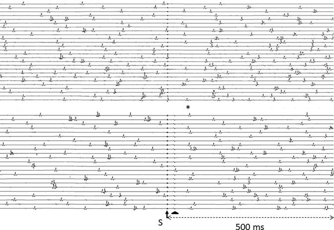
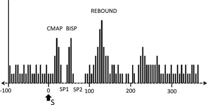
Methods: In 18 healthy volunteers, we recorded from the first dorsal interosseous (FDI) muscle the responses to CRMS of progressively increasing intensity and level of muscle contraction. The compound muscle action potential (CMAP) and the silent period (SP) were compared to those obtained with plexus, midarm and wrist stimulation. Additionally, in a smaller number of subjects, we obtained the peristimulus time histogram (psth) of single motor unit firing in the FDI, examined the effects of vibration and recorded the modulation of sustained EMG activity in muscles of the lower limbs.
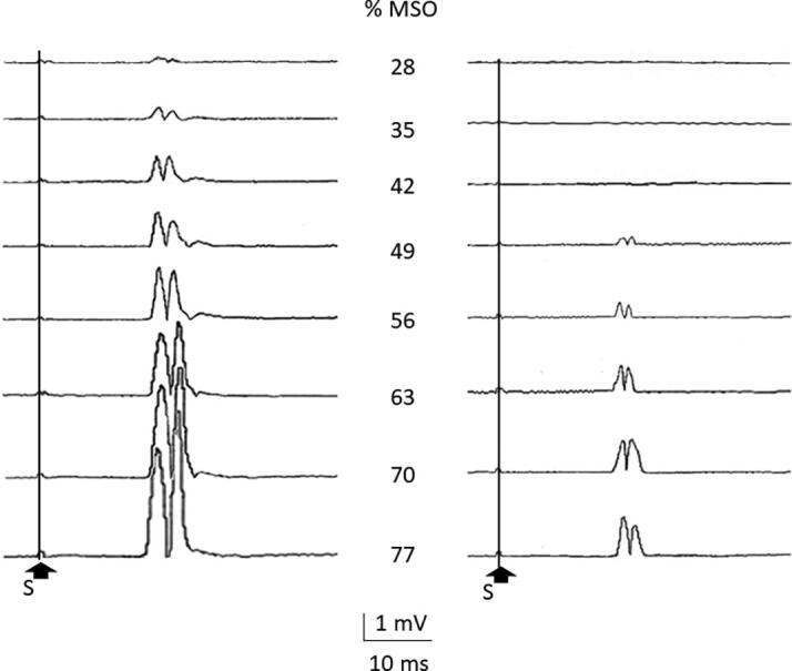
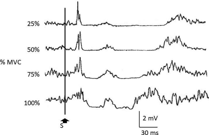
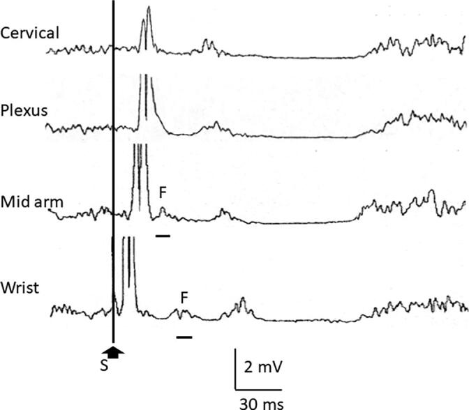
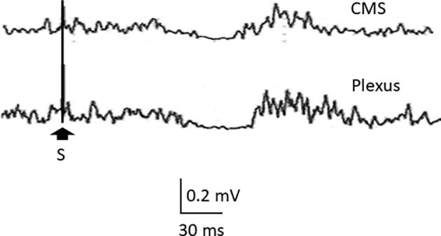
Results: Increasing CRMS intensity led to larger CMAP with no relevant changes in SP1 or SP2, except for lower amplitude of the burst interrupting the silent period (BISP). Increasing the level of muscle contraction led to reduced CMAP, shorter SP duration and increased BISP amplitude. The psth analysis showed the underlying changes in the motor unit firing frequency that corresponded to the changes seen in the CMAP and the SP with surface recordings. Progressively distal stimulation led to CMAPs of shorter latency and increased amplitude, SPs of longer latency and shorter duration, and a BISP of longer latency. Vibration led to reduction of the SP. CRMS induced SPs in muscles of the lower limb.
Conclusions: CRMS induces excitatory and inhibitory responses in hand muscles, fitting with the expected behavior of mixed nerve stimulation at very proximal sites.
Significance: Characterization of the effects of CRMS on hand muscles is of physiological and potentially clinical applicability, as it is a painless and reliable procedure.
Keywords: Cervical root magnetic stimulation; Mixed nerve stimulation; Peristimulus time histogram; Silent period.
© 2021 International Federation of Clinical Neurophysiology. Published by Elsevier B.V.
Conflict of interest statement
The authors declare that they have no known competing financial interests or personal relationships that could have appeared to influence the work reported in this paper.
Figures


Similar articles
-
Covariation between human intrinsic hand muscles of the silent periods and compound muscle action potentials evoked by magnetic brain stimulation: evidence for common inhibitory connections.Exp Brain Res. 1998 Oct;122(4):433-40. doi: 10.1007/s002210050531. Exp Brain Res. 1998. PMID: 9827862 Clinical Trial.
-
Responses of human soleus motor units to low-threshold stimulation of the tibial nerve.Exp Brain Res. 2011 Aug;213(1):73-86. doi: 10.1007/s00221-011-2779-8. Epub 2011 Jun 29. Exp Brain Res. 2011. PMID: 21713503
-
A study of synaptic connection between low threshold afferent fibres in common peroneal nerve and motoneurones in human tibialis anterior.Exp Brain Res. 2008 Dec;191(4):465-72. doi: 10.1007/s00221-008-1536-0. Epub 2008 Aug 20. Exp Brain Res. 2008. PMID: 18712371
-
Supramaximal responses can be elicited in hand muscles by magnetic stimulation of the cervical motor roots.Brain Stimul. 2010 Jul;3(3):153-60. doi: 10.1016/j.brs.2009.09.001. Epub 2009 Oct 21. Brain Stimul. 2010. PMID: 20633444
-
Silent period evoked by transcranial stimulation of the human cortex and cervicomedullary junction.J Physiol. 1993 Jul;466:521-34. J Physiol. 1993. PMID: 8410704 Free PMC article.
Cited by
-
Controversies and Clinical Applications of Non-Invasive Transspinal Magnetic Stimulation: A Critical Review and Exploratory Trial in Hereditary Spastic Paraplegia.J Clin Med. 2022 Aug 14;11(16):4748. doi: 10.3390/jcm11164748. J Clin Med. 2022. PMID: 36012986 Free PMC article.
References
-
- Ashby P. Some spinal mechanisms of negative motor phenomena in humans. Adv. Neurol. 1995;67:305–320. - PubMed
-
- Aydın Ş., Kofler M., Bakuy Y., Gündüz A., Kızıltan M.E. Effects of vibration on cutaneous silent period. Exp. Brain. Res. 2019;237(4):911–918. - PubMed
-
- Binder C., Kaya A.E., Liepert J. Vibration prolongs the cortical silent period in an antagonistic muscle. Muscle Nerve. 2009;39(6):776–780. - PubMed
-
- Burke D., Kiernan M.C., Bostock H. Excitability of human axons. Clin. Neurophysiol. 2001;112(9):1575–1585. - PubMed
LinkOut - more resources
Full Text Sources
