. 2022 Feb;48(2):e12779.
doi: 10.1111/nan.12779. Epub 2021 Dec 10.

Combinatorial model of amyloid β and tau reveals synergy between amyloid deposits and tangle formation

Affiliations

Combinatorial model of amyloid β and tau reveals synergy between amyloid deposits and tangle formation

Emily J Koller et al. Neuropathol Appl Neurobiol. 2022 Feb.

Abstract

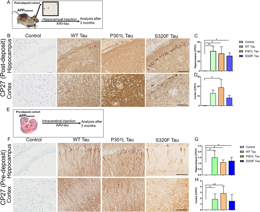
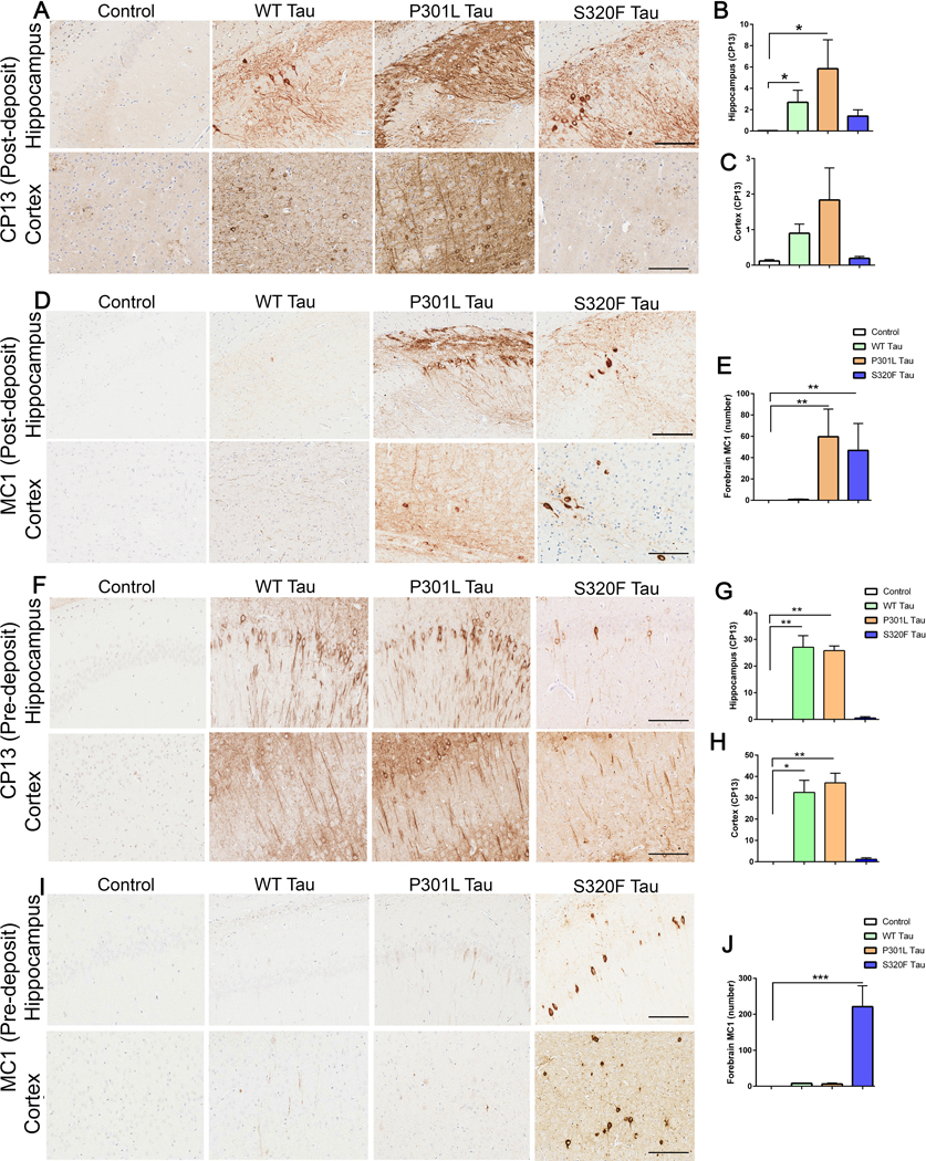
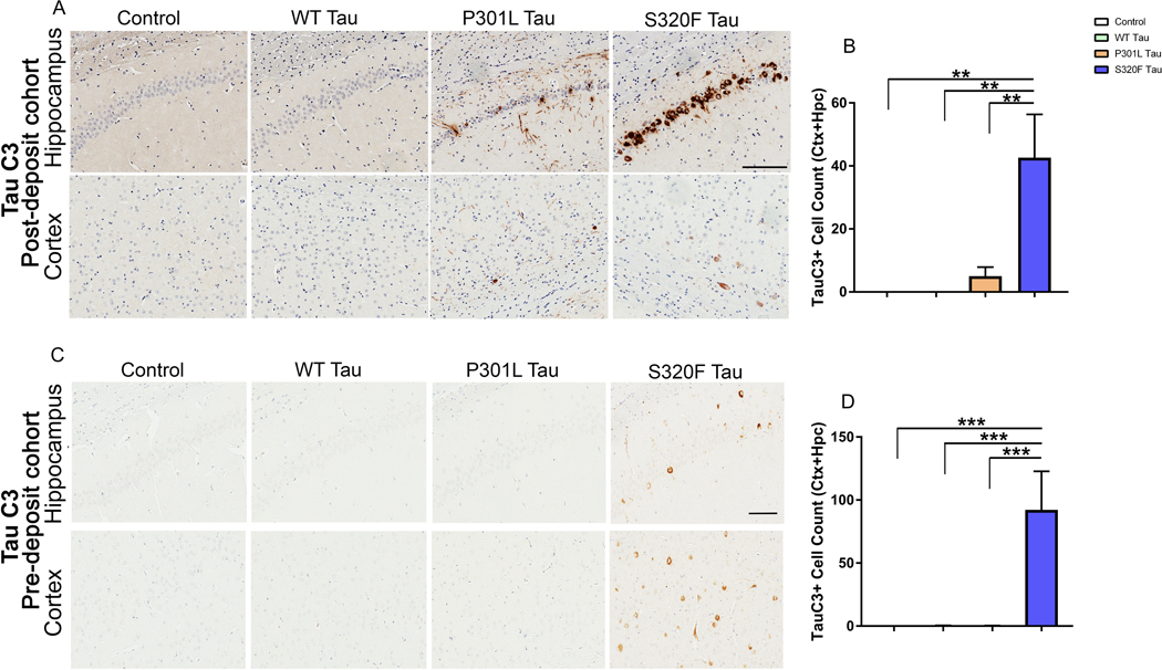
Aims: To illuminate the pathological synergy between Aβ and tau leading to emergence of neurofibrillary tangles (NFT) in Alzheimer's disease (AD), here, we have performed a comparative neuropathological study utilising three distinctive variants of human tau (WT tau, P301L mutant tau and S320F mutant tau). Previously, in non-transgenic mice, we showed that WT tau or P301L tau does not form NFT while S320F tau can spontaneously aggregate into NFT, allowing us to test the selective vulnerability of these different tau conformations to the presence of Aβ plaques.

Methods: We injected recombinant AAV-tau constructs into neonatal APP transgenic TgCRND8 mice or into 3-month-old TgCRND8 mice; both cohorts were aged 3 months post injection. This allowed us to test how different tau variants synergise with soluble forms of Aβ (pre-deposit cohort) or with frank Aβ deposits (post-deposit cohort).

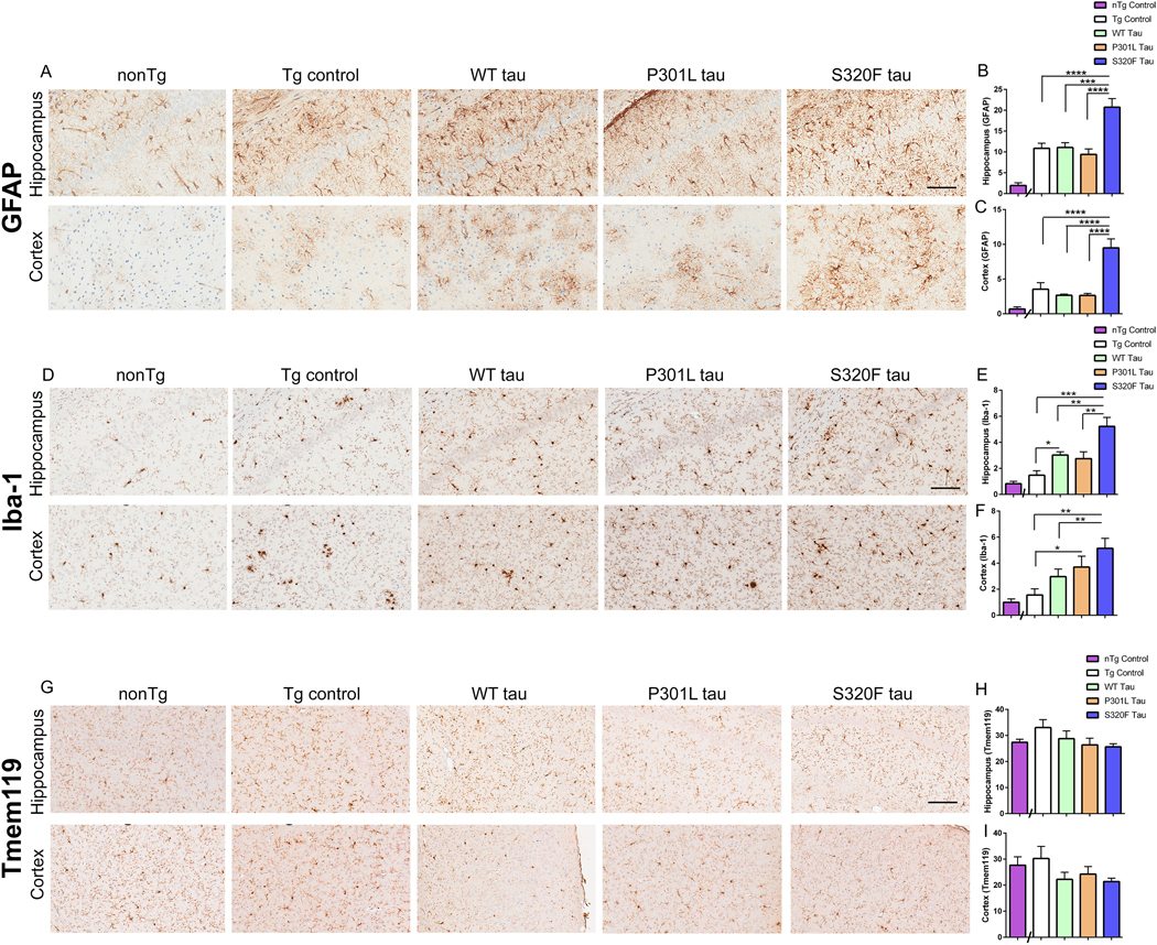
Results: Expression of WT tau did not produce NFT or altered Aβ in either cohort. In the pre-deposit cohort, S320F tau induced Aβ plaque deposition, neuroinflammation and synaptic abnormalities, suggesting that early tau tangles affect the amyloid cascade. In the post-deposit cohort, contemporaneous expression of S320F tau did not exacerbate amyloid pathology, showing a dichotomy in Aβ-tau synergy based on the nature of Aβ. P301L tau produced NFT-type inclusions in the post-deposit cohort, but not in the pre-deposit cohort, indicating pathological synergy with pre-existing Aβ deposits.

Conclusions: Our data show that different tau mutations representing specific folding variants of tau synergise with Aβ to different extents, depending on the presence of cerebral deposits.

Keywords: Alzheimer's disease; gliosis; neurofibrillary tangle; neuroinflammation; pathological synergy; plaque burden.


Conflict of interest statement

Competing interests. The authors declare that they have no conflict of interest.

Figures

Figure 1.
Figure 1.. Delivery of AAV tau variants to TgCRND8 mice results in robust human tau expression.
In the post-deposit group, 3 month old adult TgCRND8 mice were bilaterally injected into the hippocampus with AAV-WT tau, AAV-P301L tau, AAV-S320F tau, or AAV-EGFP (control) and aged to 6 months of age (A-D). In the pre-deposit group, neonatal TgCRND8 mice were injected bilaterally into the cerebral ventricles with AAV-WT tau, AAV-P301L tau, AAV-S320F tau, or AAV-EGFP (as a control) and aged to 3 months of age (E-H). Representative images of CP27 (total human tau) immunostained brain sections show tau overexpression in the hippocampus and cortex and corresponding graphs show immunoreactivity burden (mean± sem) on the y-axis. Scale bar: 100μm. n=5 mice/group. Kruskal Wallis test with Dunn’s correction; **p<0.01, *p<0.05.
Figure 2.
Figure 2.. AAV-P301L tau expression leads to tau inclusion pathology in the post-deposit group but not in the pre-deposit group.
In the post-deposit group, 3 month old adult TgCRND8 mice were bilaterally injected into the hippocampus with AAV-WT tau, AAV-P301L tau, AAV-S320F tau, or AAV-EGFP (control) and aged to 6 months of age (A-E). In the pre-deposit group, neonatal TgCRND8 mice were injected bilaterally into the cerebral ventricles with AAV-WT tau, AAV-P301L tau, AAV-S320F tau, or AAV-EGFP (as a control) and aged to 3 months of age (F-J). Representative images of CP13 (pSer202 tau) immunostained brain sections show ptau in the hippocampus and cortex of injected mice and corresponding graphs show immunoreactivity burden (mean± sem) (A-C, F-H). MC1 immunopositive inclusions were counted manually in the hippocampus and cortex (D, E, I, J). Scale bar: 100μm. n=5 mice/group. Kruskal Wallis test with Dunn’s correction;; ***p<0.001; **p<0.01, *p<0.05.
Figure 3.
Figure 3.. Induction of cleaved tau in P301L tau and S320F tau expressing post-deposit cohort mice.
Representative images of TauC3 antibody stained caspase cleaved tau is depicted in the hippocampus and cortex of post-deposit cohort (A-B) and pre-deposit cohort (C-D). The number of TauC3 positive cells were counted from each AAV-tau expressing group and presented as mean ± sem. Scale bar: 100μm. n=5 mice/group. Kruskal Wallis test with Dunn’s correction; **p<0.01, ***p<0.001.
Figure 4.
Figure 4.. Hippocampal expression of AAV-tau does not significantly alter Aβ burden in the post-deposit cohort.
Aβ plaque burden and biochemical levels of Aβ are shown for the AAV-tau or AAV-EGFP expressing post-deposit cohort (A-F) and pre-deposit cohort (G-L) of TgCRND8 mice. Representative images of 33.1.1 antibody stained Aβ plaques in the hippocampus and cortex areas and corresponding plaque burden in the whole forebrain is presented (A-B, G-H). Scale bar: 100μm. ELISA analysis of formic acid (FA) extracted detergent-insoluble Aβ42 and Aβ40 levels are depicted (C-D, I-J). ELISA analysis of detergent-soluble Aβ42 and Aβ40 levels are depicted (E-F, K-L). Data is mean± sem. Kruskal Wallis test with Dunn’s correction; **p<0.01, *p<0.05. n=5 mice/group.
Figure 5.
Figure 5.. Focused transcriptomic analysis of AAV-P301L tau expressing post-deposit cohort shows gene expression changes in immune pathways.
Relative DEG patterns of AAV-WT tau, AAV-S320F tau and AAV-P301L tau brains (normalized to AAV-EGFP) from the post-deposit group indicating changes in the different functional annotation groups indicated on the right column (A). Volcano plot (orange colour depicts upregulated genes; blue indicates downregulated genes) and corresponding DEG from AAV-WT tau mice relative to AAV-EGFP mice (B-C). Log FC denotes logarithmic value of fold change of individual genes and P-value is corrected for multiple corrections. The DEG lists top 5 upregulated and top 5 downregulated gene sorted for fold change (C). The list of upregulated and downregulated genes were used to perform a Gene ontology (GO) enrichment analysis showing enriched pathways (D, F) and a network plot highlighting connections between genes and GO terms in AAV-WT tau mice relative to AAV-EGFP (E, G). Black nodes indicate GO term “hubs” with blue & red nodes indicated down- and up-regulated gene nodes respectively. Volcano plot (H), corresponding top 10 DEG list (I), GO enriched pathways (J, L) and network analysis of GO terms (K, M) in AAV-S320F tau mice relative to AAV-EGFP mice. Volcano plot (N), corresponding top 10 DEG list (O), GO enriched pathways (P, R) and network analysis of GO terms (Q, S) in AAV-P301L tau mice relative to AAV-EGFP mice. p values adjusted for multiple testing; FDR = 0.05. Stage 1 and Stage 2 DAM score in the AAV-P301L tau mice relative to AAV-EGFP mice (T-U). 1-tailed t test; *p<0.05. n=3 mice/group.
Figure 6.
Figure 6.. AAV-P301L tau expression does not exacerbate neuroinflammation in post-deposit TgCRND8 cohort.
Representative immunohistochemical images of the hippocampus and cortex (A, D, G) and corresponding graphs depicting immunoreactivity burden (B, C, E, F, H, I) from AAV-tau and AAV-EGFP (control) expressing post-deposit TgCRND8 cohort are shown. Astrocytosis was assessed using GFAP antibody (A-C) and microgliosis assessed using Iba-1 (D-F) and Tmem119 (G-I) in the hippocampus and cortex of these mice. Data is mean± sem. Scale bar: 100μm (hippocampus), 200μm (cortex). n=5 mice/group. 1 way Anova.
Figure 7.
Figure 7.. AAV-S320F tau upregulates neuroinflammation in pre-deposit TgCRND8 cohort.
Representative immunohistochemical images of the hippocampus and cortex (A, D, G) and corresponding graphs depicting immunoreactivity burden (B, C, E, F, H, I) from AAV-tau or AAV-EGFP (control) expressing pre-deposit TgCRND8 cohort are shown. Astrocytosis was assessed using GFAP antibody (A-C) and microgliosis assessed using Iba-1 (D-F) and Tmem119 (G-I) in the hippocampus and cortex of these mice. Age-matched nonTG mice were used as baseline controls but not used in statistical analysis (n=3). Data is mean± sem. Scale bar: 100μm (hippocampus), 200μm (cortex). n=5 mice/group. 1 way Anova; ****p<0.0001, ***p<0.001, **p<0.01, *p<0.05.
Figure 8.
Figure 8.. Synaptic protein levels in AAV-tau expressing TgCRND8 mice.
Immunoblots and corresponding quantitative analysis of synaptic protein bands normalized to housekeeping gene (actin or GAPDH) are shown for AAV-tau expressing post-deposit TgCRND8 cohort (A-D) and pre-deposit TgCRND8 cohort (E-H). Molecular weight markers in kDa are shown on the left of each immunoblot. Data is mean± sem. n=3 mice/group. 1 way Anova; **p<0.01, *p<0.05.

References

    1. Hardy JA, Higgins GA. Alzheimer’s disease: the amyloid cascade hypothesis. Science. 1992;256(5054):184–5. - PubMed
    1. Karran E, De Strooper B. The amyloid cascade hypothesis: are we poised for success or failure? Journal of neurochemistry. 2016;139 Suppl 2:237–52. - PubMed
    1. Lewis J, Dickson DW, Lin WL, Chisholm L, Corral A, Jones G, et al. Enhanced neurofibrillary degeneration in transgenic mice expressing mutant tau and APP. Science. 2001;293(5534):1487–91. - PubMed
    1. Gotz J, Chen F, van Dorpe J, Nitsch RM. Formation of neurofibrillary tangles in P301l tau transgenic mice induced by Abeta 42 fibrils. Science. 2001;293(5534):1491–5. - PubMed
    1. Oddo S, Billings L, Kesslak JP, Cribbs DH, LaFerla FM. Abeta immunotherapy leads to clearance of early, but not late, hyperphosphorylated tau aggregates via the proteasome. Neuron. 2004;43(3):321–32. - PubMed

Publication types