. 2022 Jan;48(1):56-66.
doi: 10.1007/s00134-021-06562-4. Epub 2021 Nov 26.

Mechanisms of oxygenation responses to proning and recruitment in COVID-19 pneumonia

Affiliations

Mechanisms of oxygenation responses to proning and recruitment in COVID-19 pneumonia

Sandra Rossi et al. Intensive Care Med. 2022 Jan.

Abstract

Purpose: This study aimed at investigating the mechanisms underlying the oxygenation response to proning and recruitment maneuvers in coronavirus disease 2019 (COVID-19) pneumonia.

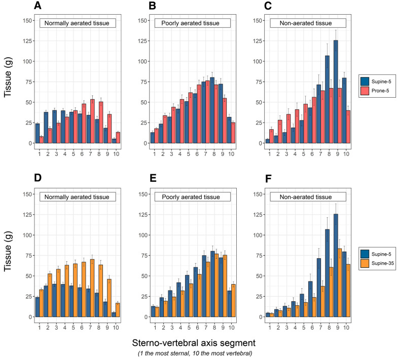
Methods: Twenty-five patients with COVID-19 pneumonia, at variable times since admission (from 1 to 3 weeks), underwent computed tomography (CT) lung scans, gas-exchange and lung-mechanics measurement in supine and prone positions at 5 cmH2O and during recruiting maneuver (supine, 35 cmH2O). Within the non-aerated tissue, we differentiated the atelectatic and consolidated tissue (recruitable and non-recruitable at 35 cmH2O of airway pressure). Positive/negative response to proning/recruitment was defined as increase/decrease of PaO2/FiO2. Apparent perfusion ratio was computed as venous admixture/non aerated tissue fraction.

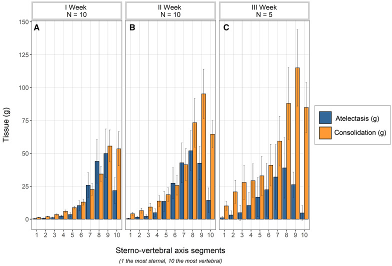
Results: The average values of venous admixture and PaO2/FiO2 ratio were similar in supine-5 and prone-5. However, the PaO2/FiO2 changes (increasing in 65% of the patients and decreasing in 35%, from supine to prone) correlated with the balance between resolution of dorsal atelectasis and formation of ventral atelectasis (p = 0.002). Dorsal consolidated tissue determined this balance, being inversely related with dorsal recruitment (p = 0.012). From supine-5 to supine-35, the apparent perfusion ratio increased from 1.38 ± 0.71 to 2.15 ± 1.15 (p = 0.004) while PaO2/FiO2 ratio increased in 52% and decreased in 48% of patients. Non-responders had consolidated tissue fraction of 0.27 ± 0.1 vs. 0.18 ± 0.1 in the responding cohort (p = 0.04). Consolidated tissue, PaCO2 and respiratory system elastance were higher in patients assessed late (all p < 0.05), suggesting, all together, "fibrotic-like" changes of the lung over time.

Conclusion: The amount of consolidated tissue was higher in patients assessed during the third week and determined the oxygenation responses following pronation and recruitment maneuvers.

Keywords: ARDS; COVID-19; Lung recruitment; Mechanical ventilation; Prone position.


Conflict of interest statement

The authors have no competing interests to disclose.

Figures

Fig. 1
Fig. 1
Upper panels: Tissue mass distributions of normally aerated (A), poorly aerated (B) and non-aerated tissues (C), as a function of lung segments (mean ± se) along the sterno (segment 1)-vertebral (segment 10) axis, in supine-5 (blue) and prone-5 positions (red). A The normally aerated tissue, in supine position, was more distributed in the ventral regions (segments 1 to 5, 179 gr ± 56, SD) and decreased in dorsal regions (segments 5 to 10, 122 gr ± 74, SD) (p < 0.001). In prone position, in contrast, it was less distributed in the ventral regions (segments 1 to 5, 122 gr ± 60, SD) and more in dorsal regions (segments 5 to 10, 200 gr ± 84, SD) (p < 0.001). B The poorly aerated tissue, in supine position, was less distributed in the ventral regions (segments 1 to 5, 161 gr ± 94, SD) and increased in dorsal regions (segments 5 to 10, 320 gr ± 112, SD) (p < 0.001). Similarly, in prone position it was less distributed in the ventral regions (segments 1 to 5, 211 gr ± 87, SD) and more in dorsal regions (segments 5 to 10, 298 gr ± 101, SD) (p < 0.001). C: the non-aerated tissue, in supine position, was markedly less distributed in the ventral regions (segments 1 to 5, 73 gr ± 92, SD) than in dorsal regions (segments 5 to 10, 427 gr ± 254, SD) (p < 0.001). Similarly, in prone position it was less distributed in the ventral regions (segments 1 to 5, 169 gr ± 154, SD) and more in dorsal regions (segments 5 to 10, 294 gr ± 209, SD) (p < 0.001). Note that differences in column heights between prone and supine from 1 to 5 indicate the formation of ventral atelectasis, while from segments 6 to 10 it indicates the disappearance of the dorsal atelectasis. Lower panels: Tissue mass distributions of normally aerated (A), poorly aerated (B) and non-aerated tissues (C), as a function of lung segments (mean ± se) along the sterno (segment 1)-vertebral (segment 10) axis, in supine-5 (blue) and supine-35 (okra yellow). D The normally aerated tissue was greater in supine-35 than in supine-5, in each of the ten segments (total normally aerated tissue 535 gr ± 171 SD vs 302 gr ± 116 SD, respectively, p < 0.001). E The poorly aerated tissue, was similar in supine-35 and in supine-5 and similarly distributed in each of the ten segments (total poorly aerated tissue 439 gr ± 189 SD vs 481 gr ± 154 SD, respectively). F: the non-aerated tissue was greater in supine-5 than in supine-35, in each of the ten segments (total non-aerated tissue 499 gr ± 328 SD vs 323 gr ± 249 SD, respectively, p < 0.05). Note that the height of the red columns represents the consolidated tissue and the difference between supine-5 and supine-35 columns represents the atelectatic tissue prevalent in the dorsal lung segments (from 5 to 10)
Fig. 2
Fig. 2
Distributions of atelectatic (blue columns) and consolidated tissue (okra yellow columns) in the ten lung segments (mean ± se) along the sterno-vertebral axis, as a function of time elapsed from admission to the study day. A First week (n = 10); B, second week (n = 10) and C, third week (n = 5). As shown, there was a significant increase of consolidated tissue overtime (p < 0.05 at the repeated measures ANOVA), while the decrease of atelectatic tissue did not reach the statistical significance

Comment in

Similar articles

Cited by

References

    1. Grasselli G, Tonetti T, Protti A, Langer T, Girardis M, Bellani G, Laffey J, Carrafiello G, Carsana L, Rizzuto C, Zanella A, Scaravilli V, Pizzilli G, Grieco DL, Di Meglio L, de Pascale G, Lanza E, Monteduro F, Zompatori M, Filippini C, Locatelli F, Cecconi M, Fumagalli R, Nava S, Vincent JL, Antonelli M, Slutsky AS, Pesenti A, Ranieri VM, Collaborators Pathophysiology of COVID-19-associated acute respiratory distress syndrome: a multicentre prospective observational study. Lancet Respir Med. 2020;8(12):1201–1208. doi: 10.1016/S2213-2600(20)30370-2. - DOI - PMC - PubMed
    1. Beloncle FM, Pavlovsky B, Desprez C, Fage N, Olivier PY, Asfar P, Richard JC, Mercat A. Recruitability and effect of PEEP in SARS-Cov-2-associated acute respiratory distress syndrome. Ann Intensive Care. 2020;10(1):55. doi: 10.1186/s13613-020-00675-7.PMID:32399901;PMCID:PMC7215140. - DOI - PMC - PubMed
    1. Habashi NM, Camporota L, Gatto LA, Nieman G. Functional pathophysiology of SARS-CoV-2-induced acute lung injury and clinical implications. J Appl Physiol (1985) 2021;130:877–891. doi: 10.1152/japplphysiol.00742.2020. - DOI - PMC - PubMed
    1. Chiumello D, Coppola S, Froio S, Mietto C, Brazzi L, Carlesso E, Gattinoni L. Time to reach a new steady state after changes of positive end expiratory pressure. Intensive Care Med. 2013;39:1377–1385. doi: 10.1007/s00134-013-2969-x. - DOI - PubMed
    1. Gattinoni L, Caironi P, Cressoni M, Chiumello D, Ranieri VM, Quintel M, Russo S, Patroniti N, Cornejo R, Bugedo G. Lung recruitment in patients with the acute respiratory distress syndrome. N Engl J Med. 2006;354:1775–1786. doi: 10.1056/NEJMoa052052. - DOI - PubMed