Review
. 2022 Apr 1;95(1132):20210217.
doi: 10.1259/bjr.20210217. Epub 2021 Dec 8.

Peritoneal malignancy: anatomy, pathophysiology and an update on modern day imaging

Affiliations
Review

Peritoneal malignancy: anatomy, pathophysiology and an update on modern day imaging

Jack W Power et al. Br J Radiol. .

Abstract

With increasing subspecialised experience in radical cytoreductive surgery and intra-abdominal chemotherapy for peritoneal malignancy, outcomes have improved significantly in selected patients. The surgery and the treatment regimens are radical and therefore correct patient selection is critical. The radiologist plays a central role in this process by estimating, as precisely as possible, the pre-treatment disease burden. Because of the nature of the disease process, accurate staging is not an easy task. Tumour deposits may be very small and in locations where they are very difficult to detect. It must be acknowledged that no form of modern day imaging has the capability of detecting the smallest peritoneal nodules, which may only be visible to direct inspection or histopathological evaluation. Nonetheless, it behoves the radiologist to be as exact and precise as possible in the reporting of this disease process. This is both to select patients who are likely to benefit from radical treatment, and just as importantly, to identify patients who are unlikely to achieve adequate cytoreductive outcomes. In this review, we outline the patterns of spread of disease and the anatomic basis for this, as well as the essential aspects of reporting abdominal studies in this patient group. We provide an evidence-based update on the relative strengths and limitations of our available multimodality imaging techniques namely CT, MRI and positron emission tomography/CT.


Figures
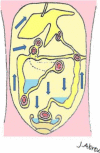
Figure 1.
Figure 1.
Pathways of ascitic fluid and potential spread of peritoneal disease. Reproduced with permission from Krishnamurthy et al.
Figure 2.
Figure 2.
In a 21-year-old patient with underlying peritoneal mesothelioma, a subtle serosal deposit at the anterior surface of the liver is seen on CT. MRI-DWI helps increase conspicuity and confidence that this is indeed a metastatic deposit. DWI, diffusion-weighted imaging.
Figure 10.
Figure 10.
A 21-year-old female with extensive peritoneal malignancy secondary to peritoneal mesothelioma. MRI-DWI images (B) show diffuse subtle deposits on the pelvic peritoneal reflections which are much more conspicuous than on the corresponding contrast enhanced CT (A). DWI, diffusion-weighted imaging.
Figure 3.
Figure 3.
A 54-year-old female with appendiceal Ca (pseudomyxoma). Here, we can see how ascitic fluid comes to lay dependently within the right and left paracolic gutters. We can also see how PET/CT can help in differentiating between reactive and malignant ascites by the presence of subtle peritoneal implants demonstrating increased metabolic activity on PET/CT. PET, positron emission tomography.
Figure 7.
Figure 7.
A 41-year-old female with diagnosis metastatic colorectal cancer. Small volume isolated abdominal wall disease implants may be difficult to identify on CT due to post-surgical changes but is well seen on PET/CT due to it’s increased metabolic activity. Although this extra-peritoneal disease makes CRS more difficult, it may be amenable to resection with the help of plastic surgeons depending on small lesion size and disease location. CRS, cytoreductive surgery; PET, positron emission tomography.
Figure 4.
Figure 4.
A 43-year-old female with peritoneal disease secondary to a moderately differentiated adenocarcinoma of colonic origin. PET/CT demonstrates metabolically active disease implants in unfavourable sites around the epigastrium, infiltrating the anterior abdominal wall and within the deep pelvis involving bowel loops. PET, positron emission tomography.
Figure 5.
Figure 5.
A 73-year-old male with diagnosis of atypical mesothelioma with peritoneal disease and mesenteric disease. While solid omental disease may be easily detected at CT and more conspicuous at PET/CT, small volume mesenteric deposits can be difficult to detect (arrows) on CT. FDG uptake within these small deposits on PET/CT increases our confidence that these represent metastatic implants. FDG, fludeoxyglucose; PET, positron emission tomography
Figure 8.
Figure 8.
A 54-year-old gentleman with a history of prior hemicolectomy for colorectal cancer re-presenting for consideration for CRS and HIPEC. CT demonstrates a small implant involving the small bowel and mesentery. Given similar density to the adjacent small bowel, this implant could easily have been missed. Due to its increased metabolic activity, it is readily identified on PET/CT. In general, when present, disease involving the small bowel is usually more diffuse than visualised on imaging and would preclude surgery. However, focal small bowel disease such as in the case may be amenable to advanced surgical treatment with CRS and HIPEC. CRS, cytoreductive surgery; HIPEC, heated intraperitoneal chemotherapy; PET, positron emission tomography.
Figure 6.
Figure 6.
PCI Score. The score from each region is added to give a total score. The central region is subdivided into regions 9–12; the upper jejenum, lower jenenum, upper ileum, lower ileum. Reproduced with permission from Fehniger et al.
Figure 9.
Figure 9.
Axial CT abdomen with i.v. + PO contrast (A) & fused axial (B) and sagittal (C) PET/CT. A 57-year-old male with history of rectal cancer with Hartmann’s resection, CRS and HIPEC. Increasing soft tissue thickening is seen at pelvic stump on CT (red arrow). FDG avid soft tissue on PET/CT with mild luminal narrowing (green arrows). Rectal stump & serosal recurrence confirmed on histology. CRS, cytoreductive surgery; HIPEC, heated intraperitoneal chemotherapy; FDG, fludeoxyglucose; PET, positron emission tomography.

References

    1. Sadeghi B, Arvieux C, Glehen O, Beaujard AC, Rivoire M, Baulieux J, et al. . Peritoneal carcinomatosis from non-gynecologic malignancies: results of the EVOCAPE 1 multicentric prospective study. Cancer 2000; 88: 358–63. doi: 10.1002/(sici)1097-0142(20000115)88:2<358::aid-cncr16>3.0.co;2-o - DOI - PubMed
    1. Verwaal VJ, van Ruth S, de Bree E, van Sloothen GW, van Tinteren H, Boot H, et al. . Randomized trial of cytoreduction and hyperthermic intraperitoneal chemotherapy versus systemic chemotherapy and palliative surgery in patients with peritoneal carcinomatosis of colorectal cancer. J Clin Oncol 2003; 21: 3737–43. doi: 10.1200/JCO.2003.04.187 - DOI - PubMed
    1. Verwaal VJ, Bruin S, Boot H, van Slooten G, van Tinteren H. 8-year follow-up of randomized trial: cytoreduction and hyperthermic intraperitoneal chemotherapy versus systemic chemotherapy in patients with peritoneal carcinomatosis of colorectal cancer. Ann Surg Oncol 2008; 15: 2426–32. doi: 10.1245/s10434-008-9966-2 - DOI - PubMed
    1. Esquivel J, Elias D, Baratti D, Kusamura S, Deraco M. Consensus statement on the loco regional treatment of colorectal cancer with peritoneal dissemination. J Surg Oncol 2008; 98: 263–7. doi: 10.1002/jso.21053 - DOI - PubMed
    1. Gill RS, Al-Adra DP, Nagendran J, Campbell S, Shi X, Haase E, et al. . Treatment of gastric cancer with peritoneal carcinomatosis by cytoreductive surgery and HIPEC: a systematic review of survival, mortality, and morbidity. J Surg Oncol 2011; 104: 692–8. doi: 10.1002/jso.22017 - DOI - PubMed

MeSH terms