. 2021 Nov 26;7(48):eabi5584.
doi: 10.1126/sciadv.abi5584. Epub 2021 Nov 26.

Toughening mechanisms for the attachment of architectured materials: The mechanics of the tendon enthesis

Affiliations

Toughening mechanisms for the attachment of architectured materials: The mechanics of the tendon enthesis

Mikhail Golman et al. Sci Adv. .

Abstract

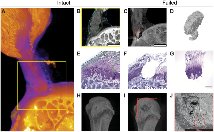
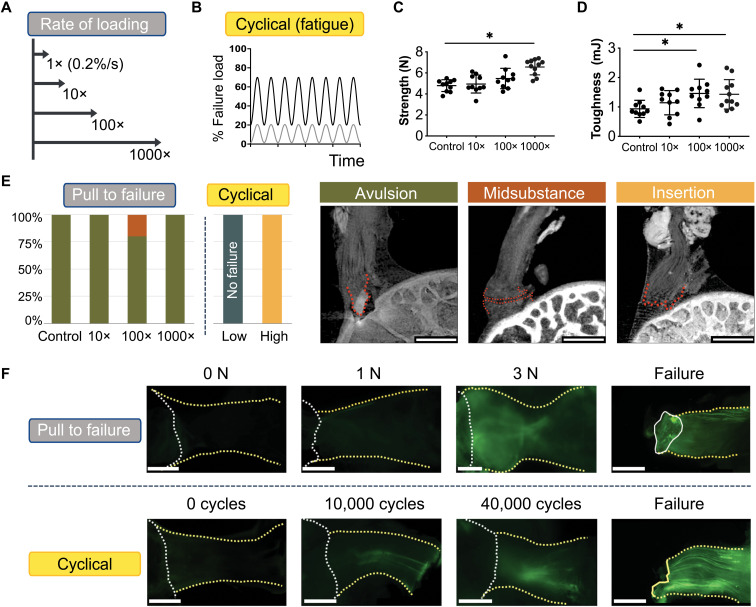
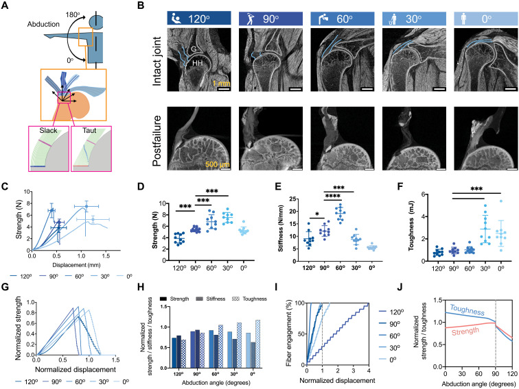
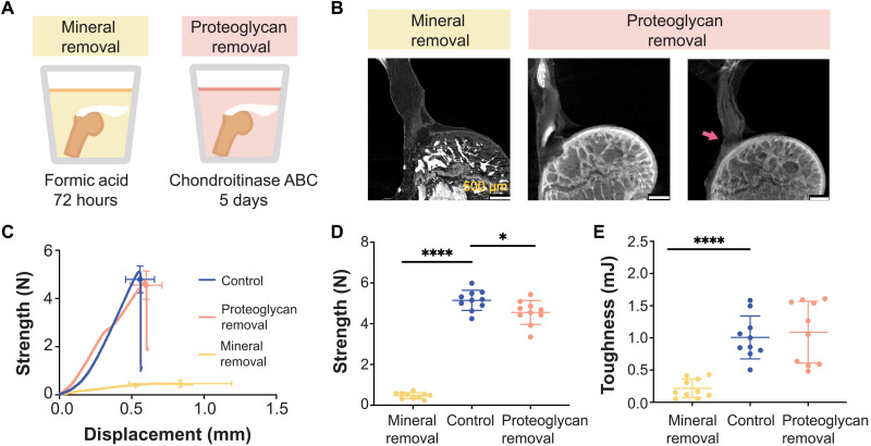
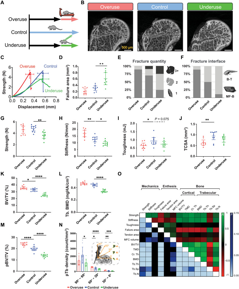
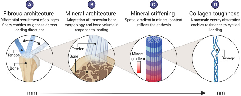
Architectured materials offer tailored mechanical properties but are limited in engineering applications due to challenges in maintaining toughness across their attachments. The enthesis connects tendon and bone, two vastly different architectured materials, and exhibits toughness across a wide range of loadings. Understanding the mechanisms by which this is achieved could inform the development of engineered attachments. Integrating experiments, simulations, and previously unexplored imaging that enabled simultaneous observation of mineralized and unmineralized tissues, we identified putative mechanisms of enthesis toughening in a mouse model and then manipulated these mechanisms via in vivo control of mineralization and architecture. Imaging uncovered a fibrous architecture within the enthesis that controls trade-offs between strength and toughness. In vivo models of pathology revealed architectural adaptations that optimize these trade-offs through cross-scale mechanisms including nanoscale protein denaturation, milliscale load-sharing, and macroscale energy absorption. Results suggest strategies for optimizing architecture for tough bimaterial attachments in medicine and engineering.


Figures

Fig. 1.
Fig. 1.. The tendon enthesis exhibits a fibrous architectured material system that fails via bony avulsion under quasi-static loading.
(A to C) HgCl2-stained contrast-enhanced high-resolution microCT imaging revealed that, hidden within the well-known larger apparent attachment footprint area is a smaller, much denser primary insertion site where tendon fibers insert directly into the bone. Imaging showed that, under quasi-static loading, only 47.4 ± 5.1% of the apparent attachment site was avulsed, revealing a previously unknown primary attachment. (A) Three-dimensional volume rendering of representative intact enthesis. (B) Magnified cross-sectional view of yellow box in (A); within blue dotted lines outline apparent enthesis and within green dotted lines outline dense primary insertion. Scale bar, 500 μm. (C) Postfailure imaging showing avulsed bony fragment at primary insertion site, outlined with a red dotted line. Scale bar, 500 μm. (D) Three-dimensional representation of avulsed fragment showing portions of trabeculae at the failure site. (E to G), Histological sections of (E) intact and (F) and (G) failed enthesis stained with toluidine blue. Scale bar, 250 μm. Blue dashed lines outline the apparent enthesis and green dotted lines outline the dense primary insertion. (H and I), Three-dimensional reconstruction from conventional microCT imaging of a representative (H) intact and (I) failed enthesis sample. (J) Scanning electron microscopy (SEM) of the failure site showing crack propagation around the avulsion site, outlined by a red circle. Scale bar, 400 μm.
Fig. 2.
Fig. 2.. Multiscale toughening mechanisms enable the entheses to exhibit distinct failure modes under varying loading conditions.
(A and B), To examine the effect of loading on failure mode, samples were loaded (A) across a range of loading rates to simulate acute injuries or (B) loaded cyclically to simulate degenerative loading. (C) Enthesis strength (i.e., failure load) and (D) enthesis toughness (i.e., energy absorption) increased with the loading rate. *P< 0.05, analysis of variance (ANOVA) followed by the Dunnet’s multiple comparison test]. (E) There were three distinct failure modes, depending on the loading regime: bone avulsion, tendon mid-substance, and tendon-bone interface (insertion failure). Scale bars, 500 μm. Under monotonic loading, most samples failed by bony avulsion failures. Under “high” cyclical loading (20 to 70% failure force), all samples failed at the insertion. Under “low” cyclical loading (1 to 20% failure force), samples did not fail, even after 100,000 cycles. (F) F-CHP fluorescence intensity, indicative of collagen damage accumulation, increased with the level of applied load and with the number of cycles. For quasi-statically loaded samples (top), there was little to no fluorescent signal in the low force group (1 to 2 N), followed by increased staining near the attachment site at higher loads (3 N and failure). For cyclically loaded samples (bottom), F-CHP staining was initially concentrated in a few fibers near the tendon mid-substance (10 to 40 K cycles) and ultimately propagated down the entire tendon in concentrated bands. Scale bars, 500 μm.
Fig. 3.
Fig. 3.. Multiscale toughening mechanisms enable the entheses to exhibit distinct failure modes under varying loading conditions.
(A) Samples were tested at varying angles of abduction (top), and a fiber recruitment model was developed to examine structural and positional contributions to enthesis toughness (bottom). (B) Contrast-enhanced microCT of intact (top row) and failed (bottom row) mouse glenohumeral joints at each abduction angle (G, glenoid; HH, humeral head). The supraspinatus tendon (top row, outlined in blue) was straight at low abduction angles (0o to 30o) and buckled at high abduction angles (90o to 120o). (C to F), There were significant differences in the attachment mechanical behavior and failure properties when samples were tested quasi-statically at varying angles ex vivo [(C) strength (failure force) versus displacement plot; (D) strength; (E) stiffness; (F) toughness] (*P < 0.05, ***P < 0.001, and ****P < 0.0001, ANOVA followed by the Dunnett’s multiple comparison test). (G to J) A positional recruitment simulation, in which fiber interactions were steric and linear, reproduced experimentally observed enthesis mechanics as a function of abduction angle. In silico (G) strength versus displacement and (H) strength, stiffness, and toughness results normalized against the case when fibers were pulled uniaxially without the geometric constraints. (I) The relationship between fiber engagement and displacement depended on abduction angle, demonstrating that the energy absorbed in reorienting and engaging fibers drove the toughening behavior of the of attachment. (J) Enthesis architecture was optimized for toughness: Normalized toughness was generally higher than normalized strength through most abduction angles.
Fig. 4.
Fig. 4.. Tendon enthesis composition drives enthesis mechanical properties.
(A) To examine compositional contributions to tendon-to-bone attachment strength and toughness, samples were immersed in decalcifying agent to completely remove mineral (left) or in chondroitinase ABC for 5 days to chemically digest proteoglycans (right). (B) Postfailure contrast-enhanced microCT scanning showed that loss of mineral or proteoglycan did not significantly alter the failure modes of the tendon enthesis. Most samples failed via bone avulsion, while a small number of samples depleted in proteoglycans failed at the edge of unmineralized fibrocartilage (pink arrow). Scale bars, 500 μm. (C to E) Quasi-static mechanical testing revealed significant differences in mechanical behavior of tendon entheses when mineral was removed. (C) Strength (failure force) versus displacement behavior. (D) Removal of mineral led to a marked decrease in strength; removal of proteoglycan led to a relatively small decrease in strength. (E) Removal of mineral led to a significant decrease in toughness; removal of proteoglycan did not affect enthesis toughness. (*P < 0.05 and ****P < 0.0001, ANOVA followed by the Dunnett’s multiple comparison test).
Fig. 5.
Fig. 5.. The tendon enthesis actively adapts its architecture in vivo by modifying mineral composition.
(A) Ten-week-old mice were subjected to two degeneration models: Underuse degeneration was induced via muscle paralysis, and overuse degeneration was achieved through downhill treadmill running for 4 weeks. (B) Postfailure contrast-enhanced microCT imaging revealed that pathological entheses exhibited exclusively avulsion-type failures under tensile mechanical testing. Scale bars, 500 μm. (C to J), Physiological in vivo degeneration models reduced the ability of the enthesis to protect against failure. (D) Failure area, (E) avulsed fragment quantity, and (F) failure interfaces were affected by enthesis pathology. Underuse degeneration led to (G) lower strength (P < 0.01) and (H) lower stiffness (P < 0.05) and (I) trended towards decreased toughness (P = 0.075) compared to that of control. Overuse degeneration decreased (J) tendon cross-sectional area (P < 0.01), (H) stiffened the enthesis (P < 0.01), and (I) significantly reduced toughness compared to control (P < 0.05). (K and L) Bone morphometric analysis revealed that underuse led to (K) reduced bone volume (BV/TV) (P < 0.0001) and (L) reduced bone mineral density (BMD) in the bone underlying the attachment (P < 0.0001). (M) The volume of load-bearing trabecular plates (pBV/TV) increased significantly (P < 0.0001) because of overuse and decreased significantly (P < 0.0001) because of underuse, with significant changes in their (N) orientations (P < 0.01, two-way ANOVA followed by Dunnet’s multiple comparison test). (O) Enthesis strength correlated with BMD (R = 0.60, P < 0.001), cortical thickness (R = 0.69, P < 0.001), and trabecular plate thickness (R = 0.44, P < 0.001). Enthesis toughness correlated with tendon cross-sectional area (R = 0.44, P < 0.01, Pearson correlation). (*P < 0.05, **P < 0.01, ***P < 0.001, and ****P < 0.0001, ANOVA followed by the Dunnett’s multiple comparison test unless otherwise reported).
Fig. 6.
Fig. 6.. The fibrous and mineral architectures of the tendon enthesis provide multiscale toughening mechanisms for a resilient attachment between tendon and bone.
Enthesis toughness is achieved over multiple length scales through unique fibrous and mineral architectures. At the millimeter-length scale, (A) the fibrous architecture of the tendon enthesis allows for fiber recruitment and reorientation to optimize toughness over strength across a range of loading directions. At the micrometer-length scale, (B) the enthesis actively adapts its mineral architecture to maintain its strength along the axis of loading. At the micrometer-to-nanometer–length scale, (C) a spatial gradient in mineral across the enthesis reduced stress concentrations (16). At the nanometer-length scale, (D) collagen damage localization protects against damage prorogating to higher length scales. Created with Biorender.com.

References

    1. Yin Z., Hannard F., Barthelat F., Impact-resistant nacre-like transparent materials. Science 364, 1260–1263 (2019). - PubMed
    1. Liu Y., Fleck N. A., Deshpande V. S., Srivastava A., High fracture toughness micro-architectured materials. J. Mech. Phys. Solids 143, 104060 (2020).
    1. Fleck N. A., Deshpande V. S., Ashby M. F., Micro-architectured materials: Past, present and future. Proc. R. Soc. A Math. Phys. Eng. Sci. 466, 2495–2516 (2010).
    1. Mirkhalaf M., Dastjerdi A. K., Barthelat F., Overcoming the brittleness of glass through bio-inspiration and micro-architecture. Nat. Commun. 5, 3166 (2014). - PubMed
    1. Barthelat F., Yin Z., Buehler M. J., Structure and mechanics of interfaces in biological materials. Nat. Rev. Mater. 1, 16007 (2016).