. 2022 May;42(5):826-843.
doi: 10.1177/0271678X211062955. Epub 2021 Nov 26.

Physical therapy exerts sub-additive and suppressive effects on intracerebral neural stem cell implantation in a rat model of stroke

Affiliations

Physical therapy exerts sub-additive and suppressive effects on intracerebral neural stem cell implantation in a rat model of stroke

Harmanvir Ghuman et al. J Cereb Blood Flow Metab. 2022 May.

Abstract

Intracerebral cell therapy (CT) is emerging as a new therapeutic paradigm for stroke. However, the impact of physical therapy (PT) on implanted cells and their ability to promote recovery remains poorly understood. To address this translational issue, a clinical-grade neural stem cell (NSC) line was implanted into peri-infarct tissue using MRI-defined injection sites, two weeks after stroke. PT in the form of aerobic exercise (AE) was administered 5 × per week post-implantation using a paradigm commonly applied in patients with stroke. A combined AE and CT exerted sub-additive therapeutic effects on sensory neglect, whereas AE suppressed CT effects on motor integration and grip strength. Behavioral testing emerged as a potentially major component for task integration. It is expected that this study will guide and inform the incorporation of PT in the design of clinical trials evaluating intraparenchymal NSCs implantation for stroke.

Keywords: Physical therapy; aerobic exercise; cell therapy; neural stem cell; neurorehabilitation; stroke.


Conflict of interest statement

Declaration of conflicting interests: The author(s) declared no potential conflicts of interest with respect to the research, authorship, and/or publication of this article.

Figures

Figure 1.
Figure 1.
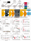
Experimental outline and behavioral analyses. (a) Sprague-Dawley rats (n = 100) were randomly allocated to experimental groups after inclusion and exclusion criteria were applied. (b) Behavioral measures, consisting of maximal capacity testing (MCT), footfault (FF) and bilateral asymmetry test (BAT) were acquired prior to middle cerebral artery occlusion (MCAo), as well as post-stroke. Two weeks post-MCAo, rats underwent T2-weighted magnetic resonance imaging (MRI) and diffusion tensor imaging (DTI) before being randomly assigned to experimental groups. Four weeks after therapeutic interventions were initiated, behavioral testing, including rotameter (Rota) and grip strength (GS), were evaluated weekly for the impact of exercise, cells and their combination onContinued.behavioral impairments. After all behavioral testing was completed 10 weeks post-implantation, a final MRI acquisition included measurement of baseline cerebral blood volume (CBV) and forepaw-stimulated functional MRI (fMRI). Animals were perfusion fixed with blood and brain being harvested for ex vivo analyses. (c) The bilateral asymmetry test (BAT) indicated an equivalent performance between all groups for tape removal of the unaffected paw. In contrast, the affected paw revealed a significant level of sensory neglect in all animals with stroke. Evaluating the experimental effect size (i.e. % change between pre- and final time point post-transplantation) revealed that Exercise (−33.67% SEM 8.77), Cells (−19.45%, SEM 14.11) and their Combination (−41.67%, SEM 7.47) reduced the impact of sensory neglect compared to the no treatment MCAo only condition. (d) Performance on motor integration, as assessed by the footfault test, revealed similar effects with Exercise (−44.65%, SEM 8.85), Cells (−57.37%, SEM 8.72) and a combined treatment (−44.43%, SEM 13.06) alleviating the impact of stroke. (e) However, amphetamine-induced asymmetry on the rotameter was not affected by the therapeutic interventions. A rotation bias occurs due to ischemic damage in the right striatum, with rats preferentially turning rightwards (clockwise) in response to a presynaptic dopamine receptor-mediated motor activity increase. (f) Grip strength measurement of the affected paw at the final time point indicated a performance of the Cell group equivalent to controls and significantly improved compared to MCAo only animals, but non-significantly different from the Exercise and Combined groups. Considering the ratio between both paws, however, indicated that the Cells and Combined group had similar grip strength that was significantly different from controls, as well as the MCAo only and the Exercise groups. (p < 0.05, p < 0.01 in comparison to MCAo).
Figure 2.
Figure 2.
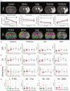
MRI-based volumetry and connectivity. a. T2-weighted (T2w) MR images reveal an extensive hyperintense region in the right hemisphere caused by stroke (10-weeks time point shown here). b. Volumetric analyses revealed a significant decrease in the ipsilateral hemisphere. Neither lesion volume, nor ipsilateral parenchymal volume changed significantly between the pre-implantation and final time point. Ventricular volume in contrast increased significantly over time. However, treatments did not affect these volumetric measurements. c. Diffusion tensor imaging (DTI) visualized the impact of stroke on brain connectivity. d. Measurements of mean diffusion (MD) revealed a significant change in the striatum due to stroke, but no effect of treatment was observed. No significant effects on MD were evident in the thalamus, motor cortex (MC) or somatosensory cortex (SMC). e. Fractional anisotropy (FA) was significantly increased after AE and Cells in the striatum and SMC compared to stroke only animals. FA in the control groups was consistently reduced at 10 weeks compared to the pre-implantation time point. f. No change in streamlines in the striatum were evident, with minor significant changes evident in the thalamus in groups receiving exercise. g. Region-to-region connectivity revealed no significant difference between groups, although exercise non-significantly increased the number of streamlines connecting the striatum with the motor (+9%) and somatosensory cortex (+15%). (p < 0.05 in comparison to MCAo).
Figure 3.
Figure 3.
Functional MRI (fMRI) and baseline cerebral blood volume (CBV). a. Group fMRI activation maps reveal brain responses to left (unaffected) or right (affected) S1 activation to forepaw stimulation (p < 0.05 voxel-wise, >80 voxels in a cluster for family-wise error correction). b. Mean activation sizes (i.e. number of active voxels) in bilateral S1 from individual rats show a significant increase in the area of activation for the cells and combined treatment group (p < 0.05 voxel-wise, >9 voxels in a cluster for family-wise correction). The response magnitudes (i.e. % fMRI signal change) and mean signal traces in affected S1 further show response recovery in the combined group. Only animals with significant activation in S1 were included in this analysis (# included/total rats); affected right S1: 8/9 control, 7/12 MCAo, 6/9 exercise, 6/8 cells, 6/9 combined; unaffected left S1: 9/9 control, 11/12 MCAo, 9/9 exercise, 6/8 cells, 7/9 combined. Threshold in A-B: p < 0.05 voxel-wise, >9 voxels in a cluster for family-wise error correction. c. Baseline fractional CBV (fCBV) maps revealed a dramatic loss of blood volume in the area of stroke. d. Quantification of fCBV in different regions of interest revealed an impact of stroke on blood volume in striatum, thalamus, motor cortex (MC), and somatosensory cortex (SMC), but no effect of treatment was evident. (p < 0.05, p < 0.01 in comparison to MCAo).
Figure 4.
Figure 4.
Graft survival. a. Neural stem cells (NSCs, STEM101+, red arrows) were implanted in the damaged peri-infarct tissue and distributed throughout this tissue, but no widespread migration was evident. The anterior-posterior image series of a rat implanted only with NSCs here illustrates the location and distribution of the graft in relation to the stroke damage. b. Nevertheless, implanted NSCs spread through several damaged tissues with very few host cells remaining. These tissues were mostly lacking host neurons (white arrow) with some implanted NSCs (red arrow) differentiating into both neurons (NeuN+/STEM101+) and astrocytes (GFAP, yellow arrow) to repopulate these regions. Very few host cells (blue arrow) were present in the graft core. c. Cell survival in the combined treatment group was reduced by 41%, but this difference did not reach statistical significance. There was no significant difference in graft volume (i.e. distribution of cells) or density of implanted cells in the tissue.
Figure 5.
Figure 5.
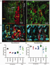
Phenotypic differentiation of implanted cells. a. Neuronal differentiation (NeuN+ cells) of CTXOE03 (STEM121+cells) was rarely observed and mostly commonly occurred in small clusters (green arrows) in the proximity of some host neurons (white arrow). b. The human-specific astrocytes marker SC123 defined the area of the grafted cells against the host background. A tight web of NSC-derived astroctytes was evident with the characteristic processes of reactive astrocytes. c. A considerable proportion of implanted human NSCs (STEM121+) co-localized with the oligodendrocyte marker SOX10 throughout the graft. A fairly homogenous distribution of SOX10 was observed, although in some instances small clusters of SOX10+ cells (green arrow) were observed in densely packed areas of the graft. d. Quantitation of differentiation indicated that <1% of grafted cells become neurons (NeuN+/STEM121+) and approximately 20% oligodendrocytes (SOX10+/STEM121+). Astrocyte (STEM123+) differentiation in the cells only group was almost 10% higher compared to the combined group.
Figure 6.
Figure 6.
Synaptogenesis and Axons. a. Synaptic density in tissues occupied by grafted cells was low and contrasted with regions containing host neurons. Nevertheless, some grafted cells were surrounded by synaptophysin (Syn)+ synapses. b. In some cases synaptophysin was evident on the cell body of a grafted cell that extended a neurofilament (NF) containing axon. c. Quantification of neurofilament revealed a significant decrease in the ipsilateral hemisphere compared to controls, but axonal density within the graft was increased in both the cell and combined groups. d. Synaptophysin was also reduced in the ischemic hemisphere compared to controls. A combined cell and exercise treatment significantly increased synapse density to control levels. Synapse density in the graft area for both the cells and combined groups was equivalent to that observed in MCAo only animals.
Figure 7.
Figure 7.
Angiogenesis and microglia response. a. Whole hemisphere images visualizing blood vessels (rat endothelial cell antigen, RECA-1), as well as microglia (Iba1) activity. b. In the graft area (SC123 marking human cells), an increased density of blood vessels was evident. c. Quantification of the area of blood vessels in the parenchyma and in the grafted area revealed a significant increase in blood vessel density in the cells and combined groups. In the graft area, the density of blood vessels was higher than in the ipsilateral hemisphere. d. Microglia density in the graft area was also increased compared to surrounding damaged tissue. e. Quantification of microglia density was increased due to MCAO, but only a localized increase in the grafted area was evident. (p < 0.05, p < 0.01, p < 0.001 in comparison to MCAo).

References

    1. Boltze J, Modo MM, Mays RW, et al.. Stem cells as an emerging paradigm in stroke 4: Advancing and accelerating preclinical research. Stroke 2019; 50: 3299–3306. - PubMed
    1. Modo MM, Jolkkonen J, Zille M, et al.. Future of animal modeling for poststroke tissue repair. Stroke 2018; 49: 1099–1106. - PMC - PubMed
    1. Smith EJ, Stroemer RP, Gorenkova N, et al.. Implantation site and lesion topology determine efficacy of a human neural stem cell line in a rat model of chronic stroke. Stem Cells 2012; 30: 785–796. - PubMed
    1. Stroemer P, Patel S, Hope A, et al.. The neural stem cell line CTX0E03 promotes behavioral recovery and endogenous neurogenesis after experimental stroke in a dose-dependent fashion. Neurorehabil Neural Repair 2009; 23: 895–909. - PubMed
    1. Hassani Z, O'Reilly J, Pearse Y, et al.. Human neural progenitor cell engraftment increases neurogenesis and microglial recruitment in the brain of rats with stroke. PLoS One 2012; 7: e50444. - PMC - PubMed

Publication types