

Tuck-and-pull technique for posterior chamber phakic intraocular lens explantation
- PMID: 34827034
- PMCID: PMC8837325
- DOI: 10.4103/ijo.IJO_652_21
Tuck-and-pull technique for posterior chamber phakic intraocular lens explantation
Abstract
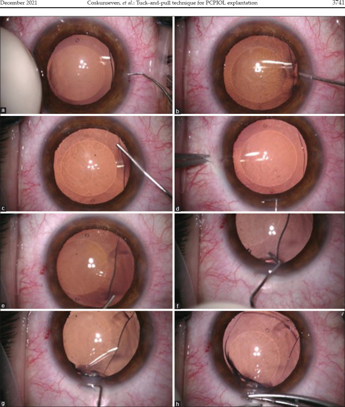
The tuck-and-pull technique was developed for practical and safe explantations of posterior chamber phakic intraocular lenses (PCPIOLs). In this technique, after the creation of a side port, viscoelastic (or OVD [ophthalmic viscosurgical device]) is initially injected behind the PCPIOL to widen the space between PCPIOL and the crystalline lens. The old incisions can be used after recent implantations rendering the enlargement of the main incision unnecessary. After additional OVD over and under the PCPIOL, the haptic is tucked by a chopper and pulled through the main incision with a single maneuver. The haptic is grasped by two suture forceps and explanted with a "hand-to-hand" maneuver. The tuck-and-pull technique provided high protection of the corneal endothelium, crystalline lens, anterior chamber structures, and the PCPIOL itself. This technique is a practical, easy, and safe approach for explantations of all PCPIOL types, whatever the reason for its explantation may be.
Keywords: Explantation; ICL; myopia; new technique; posterior chamber phakic IOL.
Conflict of interest statement
None
Figures
References
-
- Sanders DR, Vukich JA, Doney K, Gaston M. Implantable Contact Lens in Treatment of Myopia Study Group. U. S. Food and Drug Administration clinical trial of the Implantable Contact Lens for moderate to high myopia. Ophthalmology. 2003;110:255–66. - PubMed
-
- Sanders DR, Doney K, Poco M. ICL in Treatment of Myopia Study Group. United States Food and Drug Administration clinical trial of the Implantable Collamer Lens (ICL) for moderate to high myopia: Three-year follow-up. Ophthalmology. 2004;111:1683–92. - PubMed
-
- Alfonso JF, Lisa C, Fernández-Vega L, Almanzar D, Pérez-Vives C, Montés-Micó R. Prevalence of cataract after collagen copolymer phakic intraocular lens implantation for myopia, hyperopia, and astigmatism. J Cataract Refract Surg. 2015;41:800–5. - PubMed
-
- Khalifa YM, Goldsmith J, Moshirfar M. Bilateral explantation of Visian implantable collamer lenses secondary to bilateral acute angle closure resulting from a non-pupillary block mechanism. JRefract Surg. 2010;26:991–4. - PubMed
MeSH terms
LinkOut - more resources
Full Text Sources
