

Poly(lactic acid)/Zinc/Alginate Complex Material: Preparation and Antimicrobial Properties
- PMID: 34827265
- PMCID: PMC8614701
- DOI: 10.3390/antibiotics10111327
Poly(lactic acid)/Zinc/Alginate Complex Material: Preparation and Antimicrobial Properties
Abstract
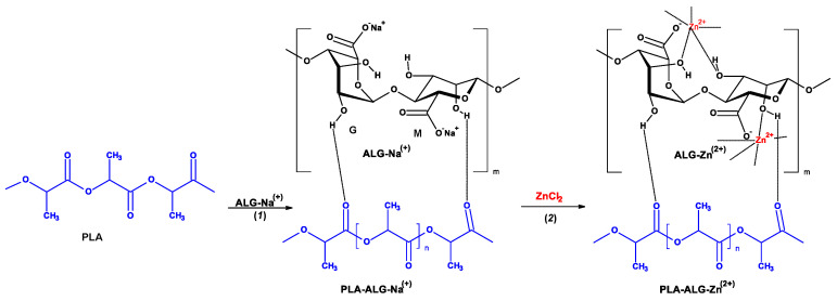
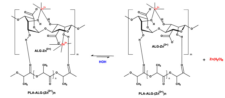
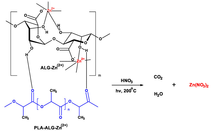
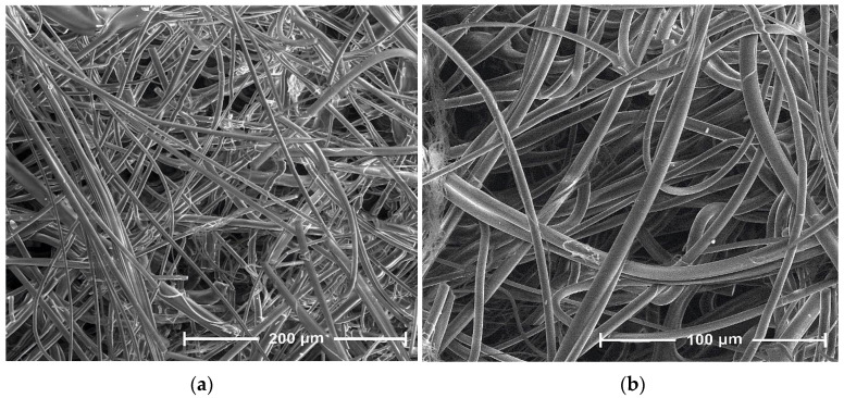
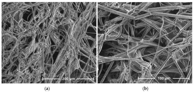
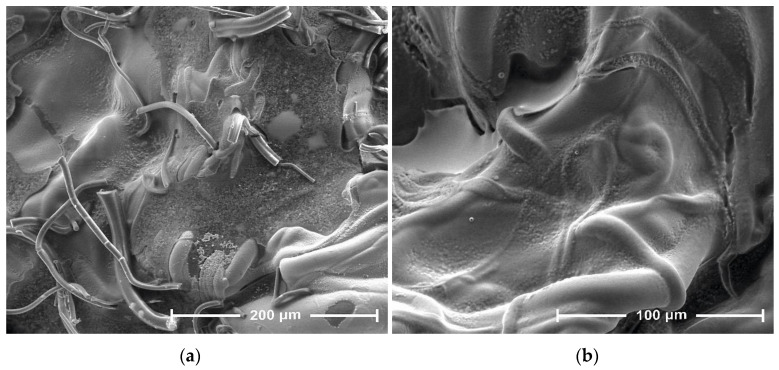
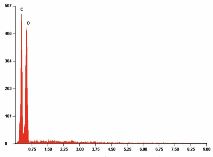
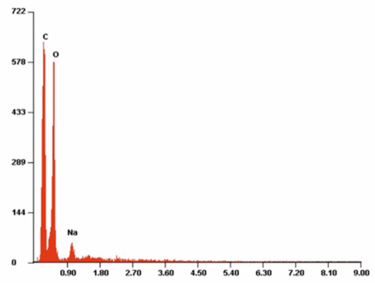
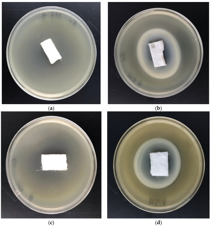
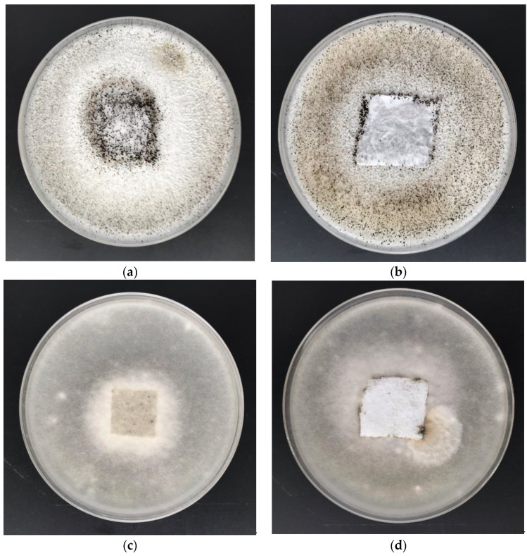
The aim of this study was to investigate an antimicrobial and degradable composite material consisting of melt-blown poly(lactic acid) nonwoven fabrics, alginate, and zinc. This paper describes the method of preparation and the characterization of the physicochemical and antimicrobial properties of the new fibrous composite material. The procedure consists of fabrication of nonwoven fabric and two steps of dip-coating modification: (1) impregnation of nonwoven samples in the solution of alginic sodium salt and (2) immersion in a solution of zinc (II) chloride. The characterization and analysis of new material included scanning electron microscopy (SEM), specific surface area (SSA), and total/average pore volume (BET). The polylactide/alginate/Zn fibrous composite were subjected to microbial activity tests against colonies of Gram-positive (Staphylococcus aureus), Gram-negative (Escherichia coli) bacterial strains, and the following fungal strains: Aspergillus niger van Tieghem and Chaetomium globosum. These results lay a technical foundation for the development and potential application of new composite as an antibacterial/antifungal material in biomedical areas.
Keywords: alginate; alginic acid; antibacterial activity; biodegradable composite; composite; melt-blown; nonwoven fabric; poly(lactide) PLA; polymer; zinc(II)chloride.
Conflict of interest statement
The authors declare no conflict of interest.
Figures
References
-
- Ghomi E.R., Khalili S., Khorasani S.N., Neisiany R.E., Ramakrishna S. Wound dressings: Current advances and future directions. J. Appl. Polym. Sci. 2019;136:47738. doi: 10.1002/app.47738. - DOI
-
- Pandey A.K., Dwivedi A.K. Recent advancement in wound healing dressing material. Int. J. Pharm. Sci. Res. 2019;10:2572–2577. doi: 10.26452/ijrps.v10i3.1512. - DOI
LinkOut - more resources
Full Text Sources
Research Materials
