Review
. 2021 Nov 6;11(11):1469.
doi: 10.3390/brainsci11111469.

Understanding Parinaud's Syndrome

Affiliations
Review

Understanding Parinaud's Syndrome

Juan Fernando Ortiz et al. Brain Sci. .

Abstract

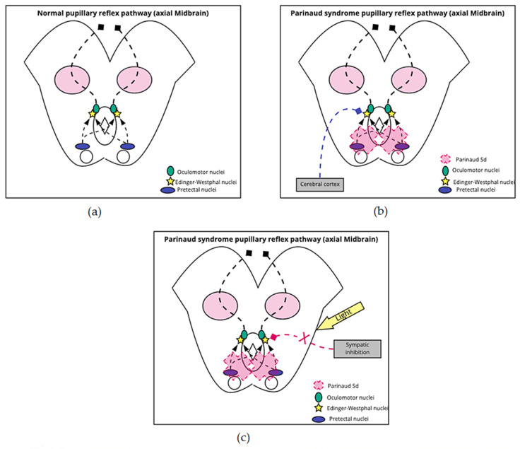
Parinaud's syndrome involves dysfunction of the structures of the dorsal midbrain. We investigated the pathophysiology related to the signs and symptoms to better understand the symptoms of Parinaud's syndrome: diplopia, blurred vision, visual field defects, ptosis, squint, and ataxia, and Parinaud's main signs of upward gaze paralysis, upper eyelid retraction, convergence retraction nystagmus (CRN), and pseudo-Argyll Robertson pupils. In upward gaze palsy, three structures are disrupted: the rostral interstitial nucleus of the medial longitudinal fasciculus (riMLF), interstitial nucleus of Cajal (iNC), and the posterior commissure. In CRN, there is a continuous discharge of the medial rectus muscle because of the lack of inhibition of supranuclear fibers. In Collier's sign, the posterior commissure and the iNC are mainly involved. In the vicinity of the iNC, there are two essential groups of cells, the M-group cells and central caudal nuclear (CCN) group cells, which are important for vertical gaze, and eyelid control. Overstimulation of the M group of cells and increased firing rate of the CCN group causing eyelid retraction. External compression of the posterior commissure, and pretectal area causes pseudo-Argyll Robertson pupils. Pseudo-Argyll Robertson pupils constrict to accommodation and have a slight response to light (miosis) as opposed to Argyll Robertson pupils were there is no response to a light stimulus. In Parinaud's syndrome patients conserve a slight response to light because an additional pathway to a pupillary light response that involves attention to a conscious bright/dark stimulus. Diplopia is mainly due to involvement of the trochlear nerve (IVth cranial nerve. Blurry vision is related to accommodation problems, while the visual field defects are a consequence of chronic papilledema that causes optic neuropathy. Ptosis in Parinaud's syndrome is caused by damage to the oculomotor nerve, mainly the levator palpebrae portion. We did not find a reasonable explanation for squint. Finally, ataxia is caused by compression of the superior cerebellar peduncle.

Keywords: collier sign; midbrain; neurology; parinaud; pseudo argyll robertson pupil.


Conflict of interest statement

The authors declare no conflict of interest.

Figures

Figure 1
Figure 1
CRN and upward gaze palsy. The superior rectus (SR) and inferior oblique muscle iOM are key for the vertical elevation of the gaze and the oculomotor (OM) nucleus. The riMLF sends information to three destinations: (1) directly to the (SR) and inferior oblique muscle (IOs), (2) to the iNC, which send their axons to the nPC (nucleus of the posterior commissure, (3) to the M group, and CCN which also innervate the RM and iOM nuclei, assisting with upward gaze. CRN is caused by a dysfunction in the riMLF and the nPC, the riMLF receives afferent projections for the center of vergence and convergence. The center of convergence, divergence is located dorsolateral to the oculomotor nerve in the dorsal midbrain.
Figure 2
Figure 2
Pseudo-Argyll Robertson Pupils. (a) Normal reflex: Signals from the optic nerve enter the pupillary reflex pathway through the pretectal nuclei, which are located anterior to the superior colliculi. The information advances bilaterally to both Edinger–Westphal nuclei, from which exit the parasympathetic fibers that accompany the NC III on its way to the eyeball; (b,c) Parinaud’s syndrome reflex: The syndrome generates damage to the pretectal nuclei, which prevents signals from reaching the Edinger–Westphal nuclei. To ensure signal arrival to these nuclei and activation of pupillary contraction, the pretectal nuclei can be bypassed and go directly from the cortex; the other possible mechanism is the antagonistic action of light on the sympathetic inhibition of the Edinger–Westphal nuclei.
Figure 3
Figure 3
Collier’s sign. The posterior commissure elicits the pupillary reflex, generates the eyes’ vertical movement, and inhibits the upper eyelid elevation pathway. Lesions in the iNC tend to alter M and CCN-group cells. The M-group and riMLF sends excitatory output to the superior rectus (SR), inferior oblique (IO) muscles, and facial nucleus (frontalis muscle), and levator palpebrae which generate eyelid elevation. When M-group cells are stimulated, the firing rate in the CCN increases, as well. Overstimulation or under inhibition of the M group is the proposed hypothesis. Supranuclear control impairment may trigger an overstimulation of the M group by increasing the CCN firing rate. Another hypothesis states that a lesion or damage in the inhibitory nPC and the iNC limits inhibition of the M group. nPC: nuclei of the posterior commissure, iNC: interstitial nucleus of Cajal, riMFL: rostral interstitial nucleus of medial longitudinal fasciculus, OM: oculomotor nuclei, SRs: paramedian nucleus, IOs: intermediate columns nucleus, CCN: central caudal nuclear.

References

    1. Ruchalski K., Hathout G.M. A Medley of Midbrain Maladies: A Brief Review of Midbrain Anatomy and Syndromology for Radiologists. Radiol. Res. Pract. 2012;2012:258524. doi: 10.1155/2012/258524. - DOI - PMC - PubMed
    1. Gandhi N., Tsehmaister-Abitbol V., Glikstein R., Torres C. Too Much on Your “Plate”? Spectrum of Pathologies Involving the Tectal Plate. Can. Assoc. Radiol. J. 2020;71:186–194. doi: 10.1177/0846537119888410. - DOI - PubMed
    1. Levidou G., Korkolopoulou P., Agrogiannis G., Paidakakos N., Bouramas D., Patsouris E. Low-grade oligodendroglioma of the pineal gland: A case report and review of the literature. Diagn. Pathol. 2010;5:59. doi: 10.1186/1746-1596-5-59. - DOI - PMC - PubMed
    1. Naqvi S., Rupareliya C., Shams A., Hameed M., Mahuwala Z., Giyanwani P.R. Pineal Gland Tumor but not Pinealoma: A Case Report. Cureus. 2017;9:e1576. doi: 10.7759/cureus.1576. - DOI - PMC - PubMed
    1. Stavale J.N., Cavalheiro S., Lamis F.C., Neto M.A.D.P. Low-Grade Oligodendroglioma of the Pineal Region: Case Report. J. Neurol. Surg. Rep. 2015;76:e55–e58. doi: 10.1055/s-0034-1396653. - DOI - PMC - PubMed

LinkOut - more resources