

A Novel Thromboplastin-Based Rat Model of Ischemic Stroke
- PMID: 34827474
- PMCID: PMC8615413
- DOI: 10.3390/brainsci11111475
A Novel Thromboplastin-Based Rat Model of Ischemic Stroke
Abstract
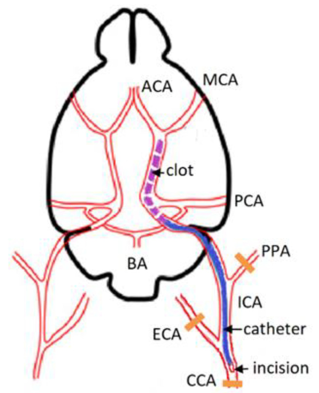
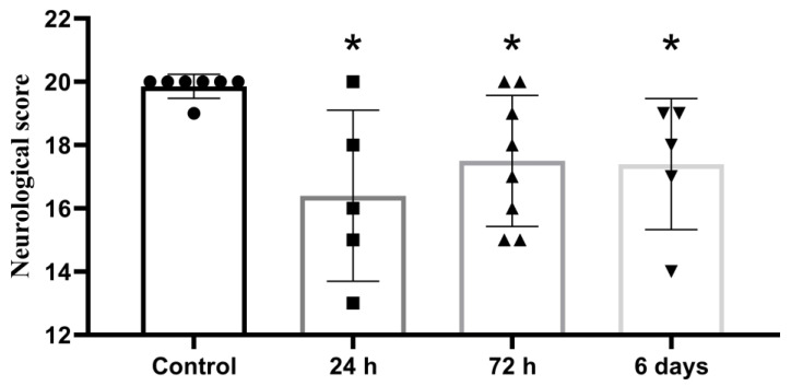
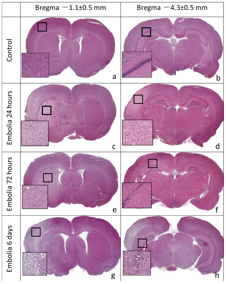
The thromboembolic ischemia model is one of the most applicable for studying ischemic stroke in humans. The aim of this study was to develop a novel thromboembolic stroke model, allowing, by affordable tools, to reproduce cerebral infarction in rats. In the experimental group, the left common carotid artery, external carotid artery, and pterygopalatine branch of maxillary artery were ligated. A blood clot that was previously formed (during a 20 min period, in a catheter and syringe, by mixing with a thromboplastin solution and CaCl2) was injected into the left internal carotid artery. After 10 min, the catheter was removed, and the incision was sutured. The neurological status of the animals was evaluated using a 20-point scale. Histological examination of brain tissue was performed 6, 24, 72 h, and 6 days post-stroke. All groups showed motor and behavioral disturbances 24 h after surgery, which persisted throughout the study period. A histological examination revealed necrotic foci of varying severity in the cortex and subcortical regions of the ipsilateral hemisphere, for all experimental groups. A decrease in the density of hippocampal pyramidal neurons was revealed. Compared with existing models, the proposed ischemic stroke model significantly reduces surgical time, does not require an expensive operating microscope, and consistently reproduces brain infarction in the area of the middle cerebral artery supply.
Keywords: brain injury; embolic stroke; hippocampus; rat model; thromboplastin.
Conflict of interest statement
The authors declare no conflict of interest.
Figures

References
-
- Chuang B.T.C., Liu X., Lundberg A.J., Toung T.J.K., Ulatowski J.A., Koehler R.C. Refinement of embolic stroke model in rats: Effect of post-embolization anesthesia duration on arterial blood pressure, cerebral edema and mortality. J. Neurosci. Methods. 2018;307:8–13. doi: 10.1016/j.jneumeth.2018.06.012. - DOI - PMC - PubMed
-
- Gafarova M.E., Naumova G.M., Gulyaev M.V., Koshelev V.B., Sokolova I.A., Domashenko M.A. Erythrocyte (dis)aggregation in stroke model in rats. Reg. Blood Circ. Microcirc. 2015;14:63–69. doi: 10.24884/1682-6655-2015-14-2-63-69. (In Russian) - DOI
LinkOut - more resources
Full Text Sources
