. 2021 Oct 31;11(11):1614.
doi: 10.3390/biom11111614.

Antioxidant and Anti-Inflammatory Effects of 3-Dehydroxyceanothetric Acid 2-Methyl Ester Isolated from Ziziphus jujuba Mill. against Cisplatin-Induced Kidney Epithelial Cell Death

Affiliations

Antioxidant and Anti-Inflammatory Effects of 3-Dehydroxyceanothetric Acid 2-Methyl Ester Isolated from Ziziphus jujuba Mill. against Cisplatin-Induced Kidney Epithelial Cell Death

Dahae Lee et al. Biomolecules. .

Abstract

Cisplatin is a platinum-based chemotherapeutic agent for treating solid tumors; however, it presents a risk factor for nephropathy. In the present study, we investigated the antioxidant and anti-inflammatory effects of 3-dehydroxyceanothetric acid 2-methyl ester (3DC2ME) isolated from Ziziphus jujuba Mill. in LLC-PK1 cells following cisplatin-induced cytotoxicity. These cells were exposed to 3DC2ME for 2 h, followed by treatment with cisplatin for 24 h. The treated cells were subjected to cell viability analysis using the Ez-Cytox assay. Reactive oxygen species (ROS) were detected via 2', 7'- dichlorodihydrofluorescein diacetate (DCFH-DA) staining. In addition, western blotting and fluorescent immunostaining were performed to evaluate protein expressions related to oxidative stress and inflammation pathways. Pretreatment with 3DC2ME protected LLC-PK1 cells from cisplatin-induced cytotoxicity and oxidative stress. In addition, pretreatment with 3DC2ME upregulated heme oxygenase 1 (HO-1) via the nuclear factor erythroid 2-related factor 2 (Nrf2) pathway in the cisplatin-treated LLC-PK1 cells. Furthermore, the increase in the expressions of IκB kinase α/β (IKKα/β), inhibitor of kappa B alpha (IκBα), nuclear factor kappa B (NF-κB), inducible nitric oxide synthase (iNOS), and cyclooxygenase-2 (COX-2) in these cells was inhibited. These results provide basic scientific evidence for understanding the antioxidant and anti-inflammatory effects of 3DC2ME isolated from Z. jujuba against cisplatin-induced kidney epithelial cell death.

Keywords: LLC-PK1; Ziziphus jujuba Mill.; inflammation; nephrotoxicity; oxidative stress.


Conflict of interest statement

The authors declare no conflict of interest.

Figures

Figure 1
Figure 1
Time-dependent cisplatin-induced oxidative stress in LLC-PK1 cells. (A) Chemical structure of cisplatin. (B) Cell viability as determined using the Ez-Cytox assay after treatment with 25 M cisplatin for 3, 6, 12, and 24 h. (C) Representative images obtained using an IX50 fluorescent microscope. The white arrows indicate dead cells (scale bar = 40 μm). (D) LLC-PK1 cells were treated with 25 M cisplatin for 3, 6, 12, and 24 h, and stained with H2DCFDA. Representative fluorescent images obtained using an IX50 fluorescent microscope. The white arrows indicate intracellular ROS (scale bar = 40 μm). (E) The fold-increases in intracellular ROS levels are represented as a bar graph. Control cells were treated with the vehicle only (mean ± SD, * p < 0.05 compared to the control).
Figure 2
Figure 2
Effect of 3DC2ME on cisplatin-induced oxidative stress in LLC-PK1 cells. (A) Chemical structure of 3DC2ME. (B) Cell viability as determined using the Ez-Cytox assay after treatment with 25 µM cisplatin in the presence or absence of 100 or 200 µM 3DC2ME for 24 h. (C) Representative images obtained using an IX50 fluorescent microscope. The white arrows indicate dead cells (scale bar = 40 μm). (D) LLC-PK1 cells exposed to 25 μM cisplatin in the presence or absence of 100 or 200 µM 3DC2ME for 24 h and stained with H2DCFDA. Representative fluorescent images were obtained using an IX50 fluorescent microscope. The white arrows indicate intracellular ROS (scale bar = 40 μm). (E) Fluorescent intensity of DCF determined using a fluorescent microplate reader. The fold-increases in intracellular reactive oxygen species (ROS) levels are represented as a bar graph. Control cells were treated with the vehicle only (mean ± SD, * p < 0.05 cisplatin-treated LLC-PK1 cells).
Figure 3
Figure 3
Effects of 3DC2ME and/or cisplatin on the expression of heme oxygenase 1 (HO-1)/nuclear factor erythroid 2-related factor 2 (Nrf2) proteins associated with antioxidant pathways in LLC-PK1 cells. (A,B) LLC-PK1 cells were exposed to 25 μM cisplatin in the presence or absence of 100 or 200 µM 3DC2ME, and western blot analysis was performed using antibodies for Nrf2, HO-1, lamin B, and GAPDH. (CE) The ratios of nuclear Nrf2, cytosolic Nrf2, and HO-1 compared with the control cells are represented as bar graphs (mean ± SD, * p < 0.05 cisplatin-treated LLC-PK1 cells).
Figure 4
Figure 4
Effects of 3DC2ME and/or cisplatin on nuclear translocation of Nrf2 in LLC-PK1 cells. LLC-PK1 cells were exposed to 25 μM cisplatin in the presence or absence of 100 or 200 µM 3DC2ME, and immunofluorescent staining was performed using antibodies for Nrf2 that were visualized with a goat anti-rabbit IgG-heavy and light chain antibody FITC conjugate (green) and mounted with ProLong® Gold antifade reagent with DAPI stained nucleus (DAPI). Representative images were visualized using an IX50 fluorescent microscope. The white arrows indicate the presence of Nrf2 (scale bar = 40 μm).
Figure 5
Figure 5
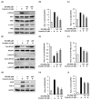
Effects of 3DC2ME and/or cisplatin on the expression of proteins associated with inflammatory pathways in LLC-PK1 cells. (A) LLC-PK1 cells were exposed to 25 μM cisplatin in the presence or absence of 100 or 200 µM 3DC2ME for 24 h, and western blot analysis was performed using antibodies for phospho-IκB kinase α/β (p-IKKα/β), IκB kinase α (IKKα), IκB kinase β (IKKβ), phospho-inhibitor of kappa B alpha (p-IκBα), inhibitor of kappa B alpha (IκBα), nuclear factor kappa B (NF-κB), and glyceraldehyde 3-phosphate dehydrogenase (GAPDH). (B,C) The ratios of p-IKKα/β and p-I-κBα compared with the control cells are represented as bar graphs. (D) LLC-PK1 cells were exposed to 25 μM cisplatin in the presence or absence of 100 or 200 µM 3DC2ME for 24 h, and western blot analysis was performed using antibodies for NF-kB, lamin B, and GAPDH. (E,F) The ratios of nuclear NF-kB and cytosolic NF-kB compared with the control cells are represented as bar graphs. (G) LLC-PK1 cells were exposed to 25 μM cisplatin in the presence or absence of 100 or 200 µM 3DC2ME for 24 h, and western blot analysis was performed using antibodies for inducible nitric oxide synthase (iNOS), cyclooxygenase-2 (COX-2), and GAPDH. (H,I) The ratios of iNOS and COX-2 compared with the control cells are represented as bar graphs (mean ± SD, * p < 0.05 cisplatin-treated LLC-PK1 cells).
Figure 6
Figure 6
Effects of 3DC2ME and/or cisplatin on nuclear translocation of NF-kB in LLC-PK1 cells. LLC-PK1 cells were exposed to 25 μM cisplatin in the presence or absence of 100 or 200 µM 3DC2ME, and immunofluorescent staining was performed using antibodies for NF-kB that was visualized with a goat anti-rabbit IgG-heavy and light chain antibody FITC conjugate (green) and mounted with ProLong® Gold antifade reagent with DAPI stained nucleus (DAPI). Representative images were obtained using an IX50 fluorescent microscope. The white arrows indicate the presence of NF-kB (scale bar = 40 μm).
Figure 7
Figure 7
Schematic pathway for the potential mechanism of 3DC2ME in renoprotection.

Similar articles

Cited by

References

    1. Lopez-Giacoman S., Madero M. Biomarkers in chronic kidney disease, from kidney function to kidney damage. World J. Nephrol. 2015;4:57. doi: 10.5527/wjn.v4.i1.57. - DOI - PMC - PubMed
    1. Pabla N., Dong Z. Cisplatin nephrotoxicity: Mechanisms and renoprotective strategies. Kidney Int. 2008;73:994–1007. doi: 10.1038/sj.ki.5002786. - DOI - PubMed
    1. Carreno J.J., Jaworski A., Kenney R.M., Davis S.L. Comparative incidence of nephrotoxicity by age group among adult patients receiving vancomycin. Infect. Dis. Ther. 2013;2:201–208. doi: 10.1007/s40121-013-0022-6. - DOI - PMC - PubMed
    1. Ramesh G., Reeves W.B. p38 MAP kinase inhibition ameliorates cisplatin nephrotoxicity in mice. Am. J. Physiol. Ren. Physiol. 2005;289:F166–F174. doi: 10.1152/ajprenal.00401.2004. - DOI - PubMed
    1. Fang C.-Y., Lou D.-Y., Zhou L.-Q., Wang J.-C., Yang B., He Q.-J., Wang J.-J., Weng Q.-J. Natural products: Potential treatments for cisplatin-induced nephrotoxicity. Acta Pharmacol. Sin. 2021 doi: 10.1038/s41401-021-00620-9. - DOI - PMC - PubMed

MeSH terms