Review
. 2021 Oct 20;12(11):1654.
doi: 10.3390/genes12111654.

Inherited Proteoglycan Biosynthesis Defects-Current Laboratory Tools and Bikunin as a Promising Blood Biomarker

Affiliations
Review

Inherited Proteoglycan Biosynthesis Defects-Current Laboratory Tools and Bikunin as a Promising Blood Biomarker

Walid Haouari et al. Genes (Basel). .

Abstract

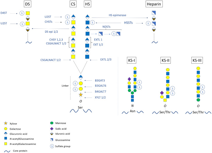
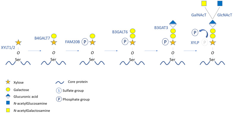
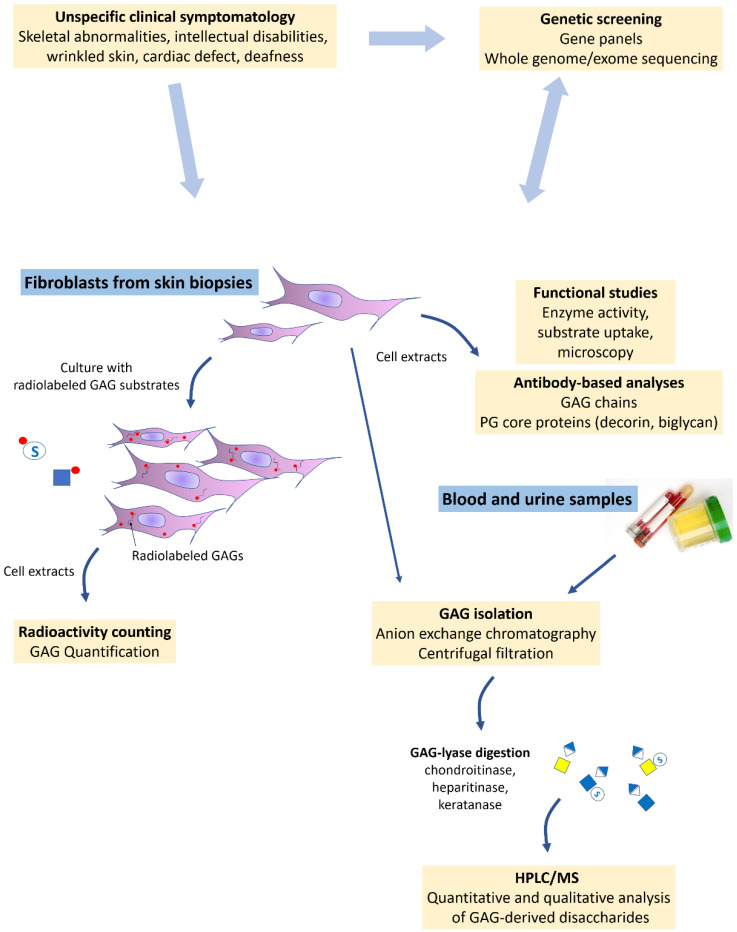
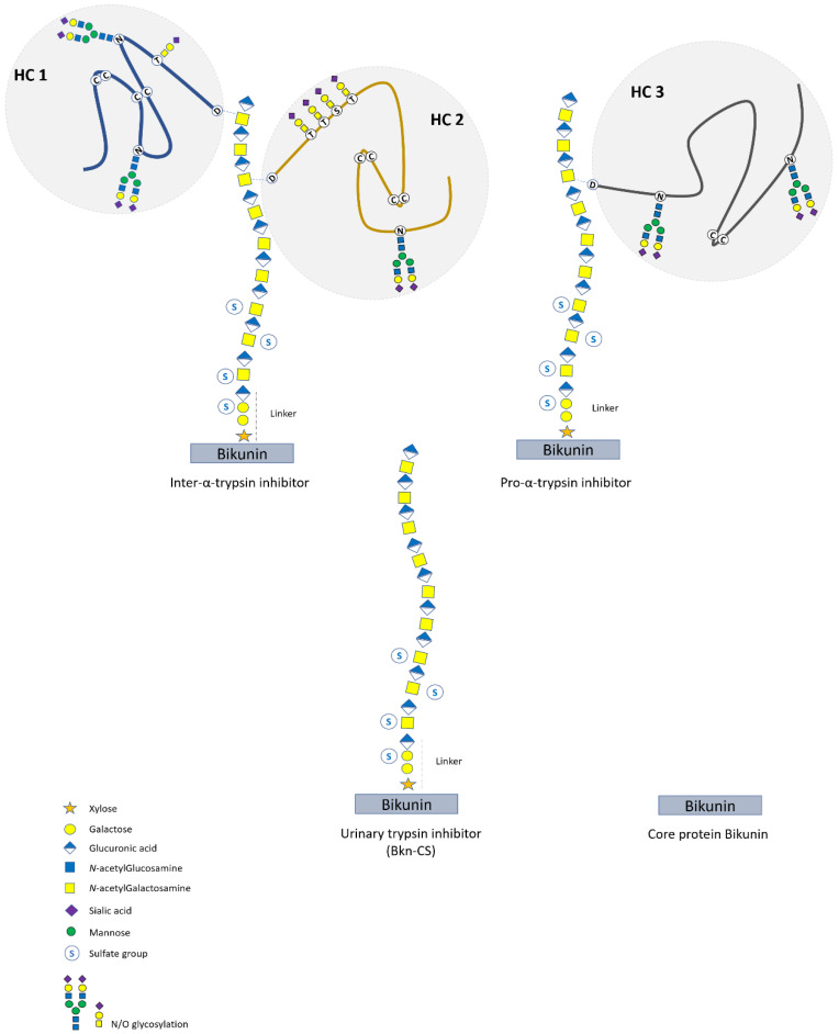
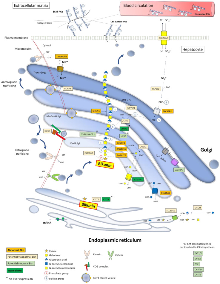
Proteoglycans consist of proteins linked to sulfated glycosaminoglycan chains. They constitute a family of macromolecules mainly involved in the architecture of organs and tissues as major components of extracellular matrices. Some proteoglycans also act as signaling molecules involved in inflammatory response as well as cell proliferation, adhesion, and differentiation. Inborn errors of proteoglycan metabolism are a group of orphan diseases with severe and irreversible skeletal abnormalities associated with multiorgan impairments. Identifying the gene variants that cause these pathologies proves to be difficult because of unspecific clinical symptoms, hardly accessible functional laboratory tests, and a lack of convenient blood biomarkers. In this review, we summarize the molecular pathways of proteoglycan biosynthesis, the associated inherited syndromes, and the related biochemical screening techniques, and we focus especially on a circulating proteoglycan called bikunin and on its potential as a new biomarker of these diseases.

Keywords: CDG; GAG; bikunin; linkeropathies; proteoglycans; skeletal dysplasia.


Conflict of interest statement

The authors declare no conflict of interest. The funders had no role in the design of the study; in the collection, analyses, or interpretation of data; in the writing of the manuscript, or in the decision to publish the results.

Figures

Figure 1
Figure 1
Biosynthesis and modifications of proteoglycans. Upon the core protein-linked tetrasaccharide linkage region (i.e., GlcA–Gal–Gal–Xyl), GAG chain polymerization starts with the transfer of a first amino sugar (i.e., GalNAc for CS and GlcNAc for HS) and continues with the addition of repetitive [GlcA–GalNAc] for CS and [GlcA–GlcNAc] for HS. Epimerization from CS to DS (i.e., [IdoA–GalNAc] backbone) and from HS to heparin (i.e., [IdoA–GlcNAc] backbone) could occur. The GAG chains are also modified by sulfate groups. Each pathway involves specific glycosyltransferases, sulfotransferases, and epimerases. For KS GAG chain elongation, there are three types of initiations leading to KS-I, KS-II, and KS-III GAG chains, which are composed of a polylactosamine backbone [GlcNAc–Gal]. Enzyme abbreviations: XYLT: Xylosyltransferase; B4GALT7: β-4-galcatosyltransferase7; B3GALT6: β-3-galactosyltransferase6; B3GAT3: β-3-glucuronyltransferase3; CSGALNACT: chondroitin sulfate N-acetylgalactosaminyltransferase; CHSY: chondroitin synthase complex; CHST: chondroitin sulfotransferase; DS epi: dermatan sulfate epimerase; U2ST: uronic acid-2-sulfotransferase; D4ST: dermatan-4-sulfotransferase; EXT: exostosin family protein; EXTL: exostosin-like; NDST: N-deacetylase/N-sulfotransferase; HSST: heparan sulfotransferase; HS epimerase: heparan sulfate epimerase.
Figure 2
Figure 2
Biosynthesis and modifications of the tetrasaccharide linkage region. The successive steps of the initiating tetrasaccharide biosynthesis and modifications are represented with the corresponding enzymes. Transfer of the first aminosugar of CS or HS chains is also illustrated. The transient phosphorylation of the xylose occurs after the first Gal addition and is removed before the first aminosugar transfer. Enzyme abbreviations: XYLT: Xylosyltransferase; B4GALT7: β-4-galactosyltransferase7; FAM20B: Family with sequence similarity 20B; B3GALT6: β-3-galactosyltransferase6; B3GAT3: β-3-glucuronyltransferase3; XYLP: xylose phosphatase; GalNAcT: N-acetylgalactosaminyltransferase; GlcNAcT: N-acetylglucosaminyltransferase.
Figure 3
Figure 3
Schematic of current PG-IMD diagnosis strategy. Once PG defect is suspected clinically, genetic screening and GAG assessment are performed using patient’s samples. Whole genome/exome sequencing allows the identification of possibly pathogenic variants in PG metabolism-associated genes. Fibroblasts from skin biopsies are cultured with radiolabeled substrates and the measured radioactivity in cell extracts reflects PG biosynthesis capacities. Fibroblasts are also used for functional analyses measuring the activity of the suspected defective enzyme or transporter, while microscopy could detect protein mislocalization and/or abnormal Golgi morphology. Antibody-based techniques such as Western blot and flow cytometry are useful for highlighting impaired PG biosynthesis by targeting GAG chains or PG core proteins such as decorin and biglycan. As from blood and urine, GAGs could be assessed by analyzing purified and enzymatically released disaccharides using HPLC/MS. Altogether, these laboratory analyses either orient towards a genetic deficiency or confirm the causality of the suspected gene variant.
Figure 4
Figure 4
Schematic structure of bikunin isoforms (IAIP). The heavy forms ITI and PαI result from the esterification of the glycoproteins HC1, HC2, and HC3 with the CS chain of the bikunin (Bkn) core protein. The light forms correspond to UTI (Bkn–CS) and the core protein Bkn. The CS chain consists of 15 +/−3 [GlcA–GalNAc] disaccharide units and is sulfated at several GalNAc residues and at the second Gal residue of the linker region.
Figure 5
Figure 5
Schematic view of Bkn–CS biosynthesis into the secretory pathway and impact of PG-IMD gene deficiencies on Bkn analysis profile. The function of each PG-IMD-associated gene is depicted together with the Bkn–CS elongation, modifications, and sorting within the secretory pathway in hepatocytes. The latter include an ER-to-Golgi anterograde vesicular flow along microtubules as ensured by dynein molecular motors while retrograde trafficking within the Golgi involves tethering factors (COG complex) and Rab-GTPase-associated proteins such as GORAB. Additionally, retrograde transport to the ER involves kinesins. Golgi homeostasis is maintained by ionic equilibrium (H+, Na+, Mn2+, Ca2+) involving ion transporters such as TMEM165. UDP-sugars and PAPS are synthesized in the cytosol by specific enzymes and their uptake within the ER/Golgi lumen is ensured by nucleotide sugar transporters and PAPS transporters, respectively. Nascent PGs are sorted in the TGN and packed into cargo vesicles addressed through kinesin-dependent post-Golgi traffic to the plasma membrane and then to the ECM and/or to blood after exocytosis. Deficiencies in the highlighted genes lead to PG-IMD syndromes, the screening of which could benefit from serum Bkn analysis. Deep and light orange boxes represent gene deficiencies leading to abnormal (already investigated) and potentially abnormal (not yet investigated) Bkn–CS biosynthesis, respectively. Deep and light green boxes include genes whose deficiency results in normal (already investigated) and potentially normal (not yet investigated) Bkn profiles, respectively.

Similar articles

Cited by

References

    1. Schaefer L., Schaefer R.M. Proteoglycans: From structural compounds to signaling molecules. Cell Tissue Res. 2010;339:237–246. doi: 10.1007/s00441-009-0821-y. - DOI - PubMed
    1. Theocharis A.D., Skandalis S.S., Gialeli C., Karamanos N.K. Extracellular matrix structure. Adv. Drug Deliv. Rev. 2016;97:4–27. doi: 10.1016/j.addr.2015.11.001. - DOI - PubMed
    1. Bartlett A.H., Park W. Proteoglycans in host-pathogen interactions: Molecular mechanisms and therapeutic implications. Expert Rev. Mol. Med. 2010;12:e5. doi: 10.1017/S1462399409001367. - DOI - PMC - PubMed
    1. Mulloy B., Rider C.C. Cytokines and proteoglycans: An introductory overview. Biochem. Soc. Trans. 2010;34:409–413. doi: 10.1042/BST0340409. - DOI - PubMed
    1. Mizumoto S., Yamada S., Sugahara K. Molecular interactions between chondroitin–dermatan sulfate and growth factors/receptors/matrix proteins. Curr. Opin. Struct. Biol. 2015;34:35–42. doi: 10.1016/j.sbi.2015.06.004. - DOI - PubMed

Publication types

MeSH terms