

Unusual Inflammatory Tinea Infections: Majocchi's Granuloma and Deep/Systemic Dermatophytosis
- PMID: 34829218
- PMCID: PMC8617809
- DOI: 10.3390/jof7110929
Unusual Inflammatory Tinea Infections: Majocchi's Granuloma and Deep/Systemic Dermatophytosis
Abstract
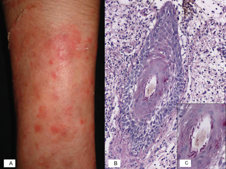
Purpose of review: Inflammatory tinea is an uncommon group of dermatophyte entities that predominantly cause fungal infection of the skin and hair. This review intends to present all of the available evidence regarding its epidemiology, etiopathogenesis, clinical features, and diagnostic methods as well as treatments recommended for various inflammatory tinea infections. This article provides a review of Majocchi's granuloma and dermatophytic or Hadida's disease.
Recent findings: The new phylogenetic classification of dermatophytes includes nine genera, and those that affect humans are Trichophyton, Microsporum, Epidermophyton, and Nannizzia. Furthermore, molecular advancements have revealed impaired antifungal immune responses caused by inflammatory tinea, which are detailed in this article.
Summary: The common denominator in these pathologies is the presence of impaired immune responses and, consequently, an impaired inflammatory response by the host. It is necessary to be familiar with these immunological characteristics in order to use the appropriate diagnostic methods and to provide adequate treatment.
Keywords: Hadida; Majocchi’s granuloma; dermatophytic; inflammatory tinea.
Conflict of interest statement
The authors declare no conflict of interest.
Figures
References
-
- Tirado-Sánchez A., Ponce-Olivera R.M., Bonifaz A. Majocchi’s Granuloma (Dermatophytic Granuloma): Updated Therapeutic Options. Curr. Fungal Infect. Rep. 2015;9:204–212. doi: 10.1007/s12281-015-0234-1. - DOI
-
- Bonifaz A., Tirado-Sánchez A., Ponce R.M. Granuloma de Majocchi. Gac. Med. Mex. 2008;144:427–433. - PubMed
Publication types
LinkOut - more resources
Full Text Sources
Research Materials
