. 2021 Nov 3;7(11):936.
doi: 10.3390/jof7110936.

Aspergillus fumigatus Fumagillin Contributes to Host Cell Damage

Affiliations

Aspergillus fumigatus Fumagillin Contributes to Host Cell Damage

Xabier Guruceaga et al. J Fungi (Basel). .

Abstract

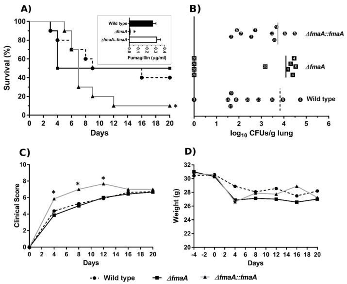
The activity of fumagillin, a mycotoxin produced by Aspergillus fumigatus, has not been studied in depth. In this study, we used a commercial fumagillin on cultures of two cell types (A549 pneumocytes and RAW 264.7 macrophages). This toxin joins its target, MetAP2 protein, inside cells and, as a result, significantly reduces the electron chain activity, the migration, and the proliferation ability on the A549 cells, or affects the viability and proliferation ability of the RAW 264.7 macrophages. However, the toxin stimulates the germination and double branch hypha production of fungal cultures, pointing out an intrinsic resistant mechanism to fumagillin of fungal strains. In this study, we also used a fumagillin non-producer A. fumigatus strain (∆fmaA) as well as its complemented strain (∆fmaA::fmaA) and we tested the fumagillin secretion of the fungal strains using an Ultra High-Performance Liquid Chromatography (UHPLC) method. Furthermore, fumagillin seems to protect the fungus against phagocytosis in vitro, and during in vivo studies using infection of immunosuppressed mice, a lower fungal burden in the lungs of mice infected with the ∆fmaA mutant was demonstrated.

Keywords: A549; Aspergillus fumigatus; RAW 264.7; UHPLC; fumagillin; mice infection; pathogenesis; virulence factor.


Conflict of interest statement

The authors declare no conflict of interest.

Figures

Figure 1
Figure 1
Fungal strains fumagillin production ability and self-resistance to the toxin. (A) Quantification of fumagillin secretion by the three A. fumigatus strains after 48 h of incubation in complete RPMI measured by UHPLC using commercial fumagillin (0.2 µg/mL) as an internal control. (* p < 0.05). (BD) Germination and double branch assay of the Wt, ΔfmaA, and ΔfmaA::fmaA, respectively, after 8 h of exposition to 0.5, 1, and 2 µg/mL of fumagillin. All the experiments were done in triplicate in three independent assays. Significant differences respect to the control without the toxin are represented (* p < 0.05, *** p < 0.001).
Figure 2
Figure 2
Quantification of the fumagillin absorption ability of each cell line. Fumagillin (1 µg/mL in complete RPMI) was added to empty wells (black line) or wells containing either A549 (dark grey) or RAW 264.7 cells (light grey), and its concentration was determined at 8, 20, and 22 h after inoculation by UHPLC. The total loss of fumagillin concentration after 24 h of exposition, compared to the initial concentration, is represented in percentage on the right axis of the graph (vertical dot lines). All the experiments were done in triplicate in three independent assays (* p < 0.05).
Figure 3
Figure 3
Fumagillin effect on A549 lung epithelial cells. (A) Electron transport chain activity of cell cultures growing in the presence of different fumagillin concentrations after 24 h of treatment. Results are expressed relative to cells growing without fumagillin. (B) Cell viability of propidium iodide-stained A549 cells measured by flow cytometry. (C) Wound closure of A549 cells when exposed to fumagillin for 24 and 48 h. (D) CFSE analysis of the A549 cell line in the presence of 0.5, 1, and 2 µg/mL of fumagillin after 48 and 72 h. Data are shown as the percentage of cells normalized versus the CFSE transferred. The grey areas represent the control cells without fumagillin, while the black line represents the cells after exposition to the mycotoxin. (E) Overlay results obtained at 72 h with the three concentrations of fumagillin tested. (F) Fluorescence intensity of the CFSE results after 48 and 72 h of treatment with fumagillin. In CFSE terms, more fluorescence means less cell proliferation. All the experiments were done by triplicate in three independent assays. (AC,F) Data were analyzed by one-way ANOVA with respect to controls without fumagillin. (* p < 0.05; *** p < 0.001).
Figure 4
Figure 4
Fumagillin effect on RAW 264.7 macrophages. (A) Cell activity of the cell cultures growing in the presence of different fumagillin concentrations for 24 h. Results are expressed relative to the cells growing without fumagillin. (B) Cell viability of propidium iodide-stained RAW 264.7 cells measured by flow cytometry. (C) CFSE analysis of RAW 264.7 cells exposed to 0.5, 1, and 2 µg/mL of fumagillin for 72 h. The percentage of cells normalized versus the CFSE transferred at 48 h and 72 h is plotted. The grey areas represent the control cells without fumagillin, while the black lines represent the results of the cells after exposition to the mycotoxin. (D) The last panel shows the overlay results obtained at 72 h with the three concentrations of fumagillin tested. (E) Fluorescence intensity of the CFSE results after 48 and 72 h of treatment with fumagillin. In CFSE terms, more fluorescence means less cell proliferation. All the experiments were done in triplicate in three independent assays. (A,B,E) Data were analyzed by one-way ANOVA respect to controls without fumagillin. (* p < 0.05; *** p < 0.001).
Figure 5
Figure 5
Growth dynamics of fungal strains in co-incubation with macrophages. Data of fungal germination (%), double branch germination (%), and hyphal length after 8 h of incubation of the three fungal strains growing either alone (A,D,G) (Control), in co-culture with BMMs (B,E,H), or in co-culture with RAW 264.7 macrophages (C,F,I). * p < 0.05.
Figure 6
Figure 6
Phagocytosis dynamics in fungus–macrophage co-cultures. (A) Phagocytosis results of co-incubation of each fungal strain and the macrophage cell line RAW 264.7. (B) Phagocytosis results of macrophages RAW 264.7 pre-treated with 1µg/mL of fumagillin for 24 h and then co-incubated with each fungal strain. After the treatment time, the RPMI medium with fumagillin was replaced with fresh complete RPMI. (C) Phagocytosis results of co-incubation between heat-inactivated conidia of the three fungal strains and RAW 264.7 cells. (D) Phagocytosis results of co-incubation of the three fungal strains and BMMs. (E) TNF production by BMMs during co-incubation with each fungal strain. All the experiments were done in triplicate in three independent assays. In each case and time point, co-cultures of Wt strain were used as control (* p < 0.05). In the case of panel (B), a solid triangle means p < 0.005 in comparison with the same time point in panel (A). Comparisons between different time points were not plotted.
Figure 7
Figure 7
Intranasal infection of a leukopenic mouse model with A. fumigatus. (A) Kaplan–Meier analysis of infected mice during 20 days post intranasal exposition to Wt, ∆fmaA, and ∆fmaA::fmaA. The panel also shows the fumagillin production ability of the conidia used to inoculate the mice. (B) Fungal burden analysis of mouse lungs. The numbers inside the symbols represent the death day of each mouse; day 20 indicates survival at the end of the study period. Vertical lines show 50% mortality in each case. (C) Values of the clinical score throughout the experiment. (D) Evolution of the mice weight during the experiment. All the experiments were done in triplicate in three independent assays. Data of Wt strains were used as control (* p < 0.05).

References

    1. Debeaupuis J.P., Sarfati J., Chazalet V., Latgé J.P. Genetic diversity among clinical and environmental isolates of Aspergillus fumigatus. Infect. Immun. 1997;65:3080–3085. doi: 10.1128/iai.65.8.3080-3085.1997. - DOI - PMC - PubMed
    1. Tekaia F., Latgé J.P. Aspergillus fumigatus: Saprophyte or pathogen? Curr. Opin. Microbiol. 2005;8:385–392. doi: 10.1016/j.mib.2005.06.017. - DOI - PubMed
    1. Brakhage A.A., Langfelder K. Menacing mold: The molecular biology of Aspergillus fumigatus. Annu. Rev. Microbiol. 2002;56:433–455. doi: 10.1146/annurev.micro.56.012302.160625. - DOI - PubMed
    1. Kwon-Chung K.J., Sugui J.A. Aspergillus fumigatus-What makes the species a ubiquitous human fungal pathogen? PLoS Pathog. 2013;9:e1003743. doi: 10.1371/journal.ppat.1003743. - DOI - PMC - PubMed
    1. Schmitt H.J., Blevins A., Sobeck K., Armstrong D. Aspergillus species from hospital air and from patients. Mycoses. 1991;33:539–541. doi: 10.1111/myc.1990.33.11-12.539. - DOI - PubMed

LinkOut - more resources