Review
. 2021 Nov 22;7(11):996.
doi: 10.3390/jof7110996.

Fungal Endophthalmitis: A Comprehensive Review

Affiliations
Review

Fungal Endophthalmitis: A Comprehensive Review

Abid A Haseeb et al. J Fungi (Basel). .

Abstract

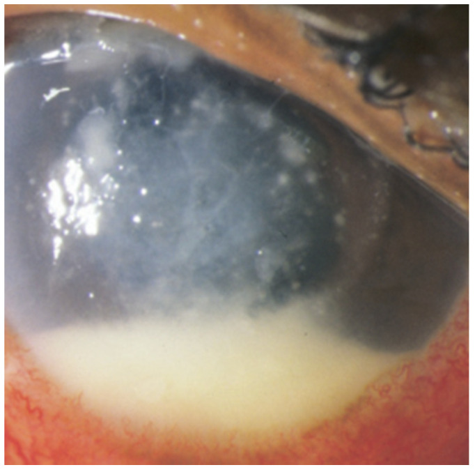
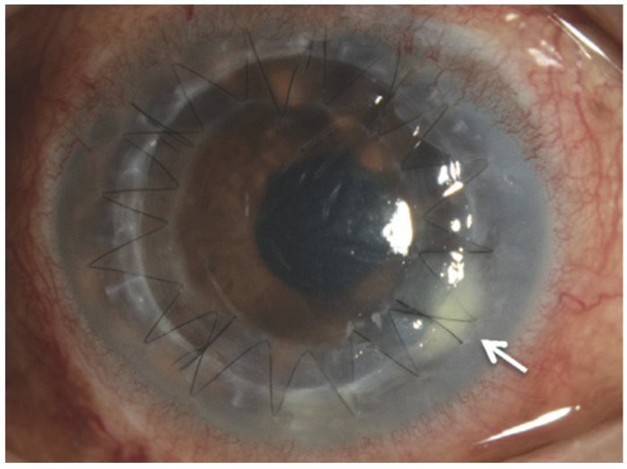
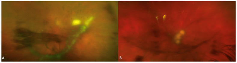
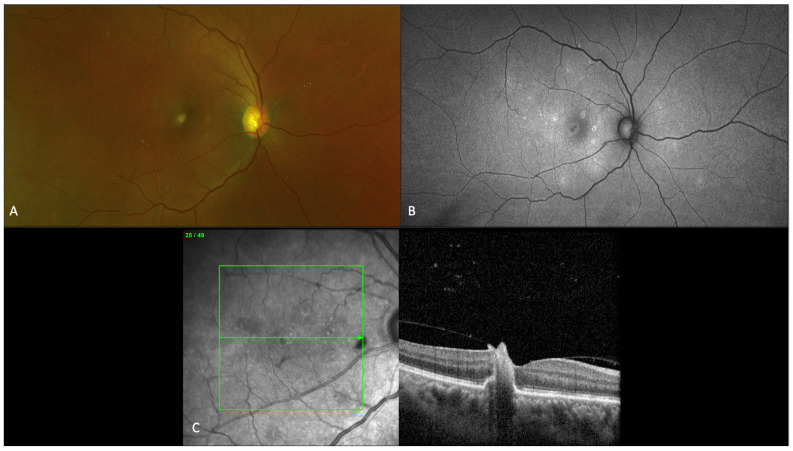
Endophthalmitis is a serious ophthalmologic condition involving purulent inflammation of the intraocular spaces. The underlying etiology of infectious endophthalmitis is typically bacterial or fungal. The mechanism of entry into the eye is either exogenous, involving seeding of an infectious source from outside the eye (e.g., trauma or surgical complications), or endogenous, involving transit of an infectious source to the eye via the bloodstream. The most common organism for fungal endophthalmitis is Candida albicans. The most common clinical manifestation of fungal endophthalmitis is vision loss, but other signs of inflammation and infection are frequently present. Fungal endophthalmitis is a clinical diagnosis, which can be supported by vitreous, aqueous, or blood cultures. Treatment involves systemic and intravitreal antifungal medications as well as possible pars plana vitrectomy. In this review, we examine these essential elements of understanding fungal endophthalmitis as a clinically relevant entity, which threatens patients' vision.

Keywords: Aspergillus; Candida; antifungals; endogenous endophthalmitis; exogenous endophthalmitis; fungal endophthalmitis; pars plana vitrectomy.


Conflict of interest statement

The authors declare no conflict of interest.

Figures

Figure 1
Figure 1
Photograph demonstrating Fusarium oxysporum endophthalmitis which developed 27 days after minor trauma with organic matter to the right eye. A hypopyon and anterior chamber fungal infiltrates are seen in the setting of a multifocal, feathery-edged corneal infiltrate. Adapted from Wykoff C.C., Flynn H.W., Jr., Miller D., Scott I.U., and Alfonso E.C. Exogenous fungal endophthalmitis: microbiology and clinical outcomes. Ophthalmology. 2008; 115(9): 1501–1507.e15072 [17]. Figure 1A, Copyright (2008) with permission from Elsevier. License 5138360747040 on 29 August 2021.
Figure 2
Figure 2
Slit-lamp examination showing that small white infiltrates were observed at the border between the host and donor corneal graft. Adapted from Kitazawa K., Wakimasu K., Yoneda K., Iliakis B., Sotozono C., and Kinoshita S. A case of fungal keratitis and endophthalmitis post penetrating keratoplasty resulting from fungal contamination of the donor cornea. Am J Ophthalmol Case Rep. 2016; 5: 103–106 [32]. Figure 2A, Copyright (2016) with permission from Elsevier. License 5135721109911 on 25 August 2021.
Figure 3
Figure 3
Fundus photographs of two patients with fungal endophthalmitis with (A) characteristic creamy-white and (B) well circumscribed “string of pearls” retinal and vitreous opacities.
Figure 4
Figure 4
Imaging findings for a 56-year-old patient with fungal chorioretinitis, showing characteristic fundus, autofluorescence, and optical coherence tomography (OCT) macula findings. (A) Fundus photograph of right eye showing multiple small chorioretinal white lesions predominantly in the macula. (B) Fundus autofluorescence showing large area of hypoautofluoroescence in the fovea and multiple areas of scattered punctate hyperautofluoresence lesions in areas of early retinal pigment epithelium loss. (C) OCT macula findings over the foveal chorioretinal lesion depicting infiltration from choroid through retinal layers into the vitreous with focal areas of traction.
Figure 5
Figure 5
Ophthalmic B-scan showing (A) an endophytic lesion on optic nerve head and vitreous opacification (B) intra-operative view during surgical removal of large endophytic fungal ball that was previously identified on B-scan.
Figure 6
Figure 6
Algorithm for medical management of fungal endophthalmitis. Abbreviations: IVT, intravitreal; PO, oral; IV, intravenous. Systemic fluconazole: 800 mg (12 mg/kg) then 400–800 mg (6–12 mg/kg) daily. Systemic voriconazole: 400 mg (6 mg/kg) intravenous twice daily for 2 doses then 300 mg (4 mg/kg) intravenous or oral twice daily. Liposomal amphotericin B: 3–5 mg/kg intravenously daily, with or without oral flucytosine 25 mg/kg four times daily. Intravitreal amphotericin B: 5–10 µg/0.1 mL. Intravitreal voriconazole: 100 µg/0.1 mL.

References

    1. Durand M.L. Endophthalmitis. Clin. Microbiol. Infect. 2013;19:227–234. doi: 10.1111/1469-0691.12118. - DOI - PMC - PubMed
    1. Durand M.L. Bacterial and Fungal Endophthalmitis. Clin. Microbiol. Rev. 2017;30:597–613. doi: 10.1128/CMR.00113-16. - DOI - PMC - PubMed
    1. Connell P.P., O’Neill E.C., Fabinyi D., Islam F.M., Buttery R., McCombe M., Essex R.W., Roufail E., Clark B., Chiu D., et al. Endogenous endophthalmitis: 10-year experience at a tertiary referral centre. Eye. 2011;25:66–72. doi: 10.1038/eye.2010.145. - DOI - PMC - PubMed
    1. Kernt M., Kampik A. Endophthalmitis: Pathogenesis, clinical presentation, management, and perspectives. Clin. Ophthalmol. 2010;4:121–135. doi: 10.2147/OPTH.S6461. - DOI - PMC - PubMed
    1. Pflugfelder S.C., Flynn H.W., Jr., Zwickey T.A., Forster R.K., Tsiligianni A., Culbertson W.W., Mandelbaum S. Exogenous fungal endophthalmitis. Ophthalmology. 1988;95:19–30. doi: 10.1016/S0161-6420(88)33229-X. - DOI - PubMed

LinkOut - more resources