. 2021 Oct 27;11(11):1999.
doi: 10.3390/diagnostics11111999.

Comparative Analysis of Circulating Biomarkers for Patients Undergoing Resection of Colorectal Liver Metastases

Affiliations

Comparative Analysis of Circulating Biomarkers for Patients Undergoing Resection of Colorectal Liver Metastases

Sven H Loosen et al. Diagnostics (Basel). .

Abstract

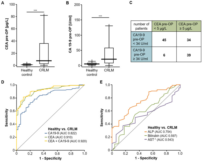
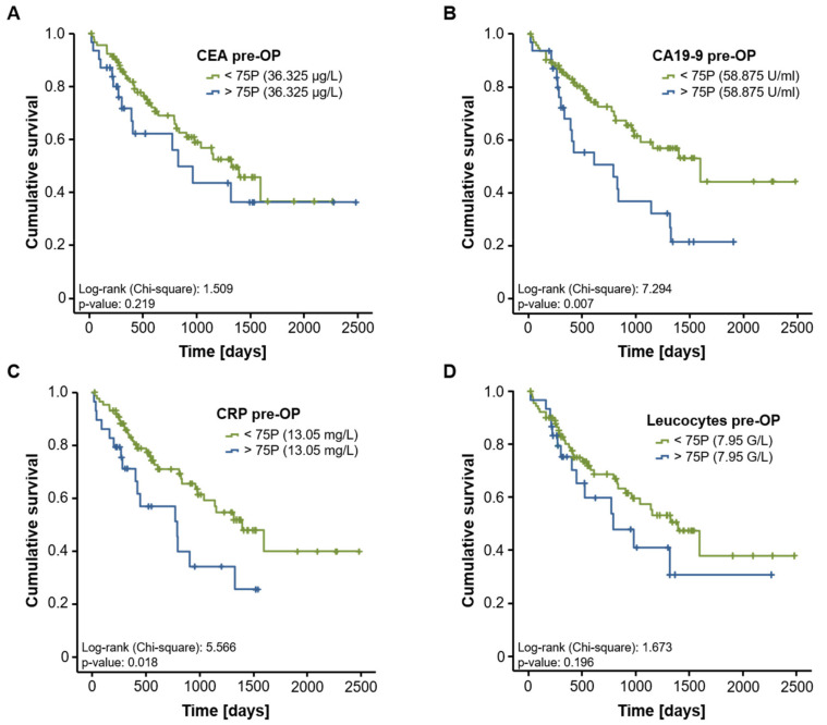
Surgical tumor resection has evolved as a potentially curative therapy for patients with resectable colorectal liver metastases (CRLM). However, disease recurrence is common and the available preoperative stratification strategies are often imprecise to identify the ideal candidates for surgical treatment, resulting in a postoperative 5-year survival rate below 50%. Data on the prognostic value of CEA, CA19-9 and other common laboratory parameters after CRLM resection are scarce and partly inconclusive. Here, we analyzed the prognostic potential of circulating CEA and CA19-9 in comparison to other standard laboratory markers in resectable CRLM patients. Serum levels of tumor markers and other laboratory parameters were analyzed in 125 patients with CRLM undergoing tumor resection at a tertiary referral center. Results were correlated with clinical data and outcome. Both tumor markers were significantly elevated in CRLM patients compared to healthy controls. Interestingly, elevated levels of CEA, CA19-9 and C-reactive protein (CRP) were associated with an unfavorable prognosis after CRLM resection in Kaplan-Meier curve analysis. However, only CEA and not CA19-9 or CRP serum levels were an independent prognostic marker in multivariate Cox regression analysis. Our data demonstrate that circulating levels of CEA rather than CA19-9 might be a valuable addition to the existing preoperative stratification algorithms to identify patients with a poor prognosis after CRLM resection.

Keywords: CA19-9; CEA; CRC; CRLM; CRP; cancer; liver resection; survival.


Conflict of interest statement

The authors declare no conflict of interest.

Figures

Figure 1
Figure 1
Serum levels of CEA and CA19-9 are elevated in patients with CRLM. Preoperative serum levels of CEA (A) and CA19-9 (B) are significantly elevated in patients with CRLM compared to healthy controls. (C) Number of patients with elevated CEA and CA19-9 levels above the standard cut-off value. (D) ROC curve analysis reveals AUC values of 0.910 and 0.822 for CEA and CA19-9, respectively, for the differentiation between CRLM patients and healthy controls. (E) Other routinely tested serum markers of liver injury have an inferior AUC. *** p < 0.001.
Figure 2
Figure 2
CEA, CA19-9 and CRP serum levels correlate with the size of CRLM. Serum levels of CEA (A), CA19-9 (B) and CRP (C) show a strong correlation with the size of CRLM. The leucocyte count does not significantly correlate with the CRLM size (D).
Figure 3
Figure 3
Evaluation of CEA, CA19-9, CRP and leucocyte count as prognostic marker after CRLM resection. Kaplan–Meier curve analysis reveals that only CA19-9 (B) and CRP (C), but not CEA (A) or leucocyte count (D), indicate an impaired long-term survival in patients with circulating levels above the 75th percentile.
Figure 4
Figure 4
CEA, CA19-9 and CRP are prognostic factors of overall survival after resection of CRLM. When using the optimal prognostic cut-off value, Kaplan–Meier curve analyses show a significant impaired long-term survival for CRLM patients with CEA serum levels above 24.55 µg/L (A), CA19-9 serum levels above 30.25 U/mL (B) and CRP levels above 6.95 mg/L (C). The ideal cut-off value for the leucocyte count (5.95 G/L) is unable to discriminate between long-term survivors and non-survivors (D).

Similar articles

Cited by

References

    1. Brenner H., Kloor M., Pox C.P. Colorectal cancer. Lancet. 2014;383:1490–1502. doi: 10.1016/S0140-6736(13)61649-9. - DOI - PubMed
    1. Jones R.P., Kokudo N., Folprecht G., Mise Y., Unno M., Malik H.Z., Fenwick S.W., Poston G.J. Colorectal Liver Metastases: A Critical Review of State of the Art. Liver Cancer. 2017;6:66–71. doi: 10.1159/000449348. - DOI - PMC - PubMed
    1. Mody K., Bekaii-Saab T. Clinical Trials and Progress in Metastatic Colon Cancer. Surg. Oncol. Clin. 2018;27:349–365. doi: 10.1016/j.soc.2017.11.008. - DOI - PubMed
    1. Van Cutsem E., Cervantes A., Adam R., Sobrero A., Van Krieken J.H., Aderka D., Aguilar E.A., Bardelli A., Benson A., Bodoky G., et al. ESMO consensus guidelines for the management of patients with metastatic colorectal cancer. Ann. Oncol. 2016;27:1386–1422. doi: 10.1093/annonc/mdw235. - DOI - PubMed
    1. Jones R.P., Jackson R., Dunne D.F.J., Malik H.Z., Fenwick S.W., Poston G.J., Ghaneh P. Systematic review and meta-analysis of follow-up after hepatectomy for colorectal liver metastases. Br. J. Surg. 2012;99:477–486. doi: 10.1002/bjs.8667. - DOI - PubMed