

Case Reports
doi: 10.3390/diagnostics11112050.
Maxillary Mucormycosis Osteomyelitis in Post COVID-19 Patients: A Series of Fourteen Cases
Affiliations
- PMID: 34829397
- PMCID: PMC8624954
- DOI: 10.3390/diagnostics11112050
Item in Clipboard
Case Reports
Maxillary Mucormycosis Osteomyelitis in Post COVID-19 Patients: A Series of Fourteen Cases
Diagnostics (Basel).
.
Abstract
During the current pandemic of COVID-19, numerous manifestations and complications have developed. Patients with COVID-19 are at high risk of fungal infections, such as mucormycosis, that may result directly from COVID-19 infection and/or as a side effect of the drugs used in COVID-19 treatment protocol, such as dexamethasone, hydroxychloroquine, and antibiotics. In this report, we described a series of 14 cases with maxillary mucormycosis osteomyelitis in immediate post-COVID-19 patients.
Keywords: COVID-19; maxilla; mucormycosis; osteomyelitis.
Conflict of interest statement
The authors declared no conflict of interest.
Figures
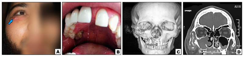
Case#2: (A) Extraoral swelling of the right side of the face (note: sinus tract inferolaterally to the eye {arrow}). (B) Exposed necrotic bone in the right posterior palate. (C,D) 3D and cronal CT showing bone destruction of right maxilla, inferior orbital rim, and lower part of zygoma.
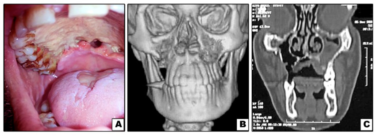
Case#3: (A) Exposed necrotic palatal bone with palatal perforations. (B,C) 3D and coronal CT showing bone destruction of right and left maxillae (not involving inferior orbital rims).
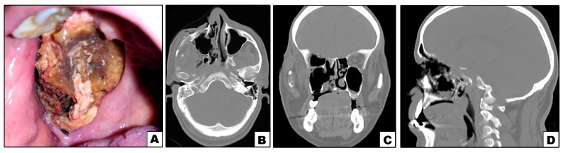
Case#5: (A) Exposed necrotic palatal and alveolar bone on right side. (B–D) Axial, coronal and sagittal CT showing bone destruction involving right maxilla and orbit.
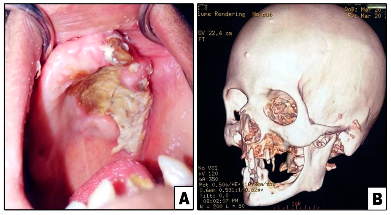
Case#7: (A) Exposed necrotic palatal and alveolar bone on left side. (B) 3D CT showing bone destruction involving left maxilla and lower part of zygoma.
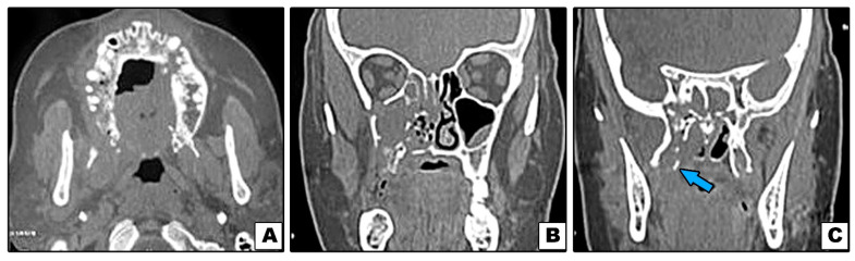
Case#13: (A–C) Axial and coronal CT showing bone destruction involving premaxilla, right maxilla with pterygoid plates (arrow), and not involving inferior orbital rim.
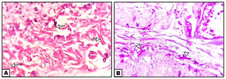
Nonseptate hyphae with right angle branching (arrows) and spores of mucormycosis surrounded by necrotic tissues and a dense inflammatory infiltration. ((A): H&E ×40, (B): PAS ×40).
References
-
- Kaye A.D., Cornett E.M., Brondeel K.C., Lerner Z.I., Knight H.E., Erwin A., Charipova K., Gress K.L., Urits I., Urman R.D., et al. Biology of COVID-19 and related viruses: Epidemiology, signs, symptoms, diagnosis, and treatment. Best Pract. Res. Clin. Anaesthesiol. 2020;35:269–292. doi: 10.1016/j.bpa.2020.12.003. - DOI - PMC - PubMed
Publication types
LinkOut - more resources
Full Text Sources
