

New Evaluation Method for Bone Formation around a Fully Hydroxyapatite-Coated Stem Using Digital Tomosynthesis: A Retrospective Cross-Sectional Study
- PMID: 34829440
- PMCID: PMC8623614
- DOI: 10.3390/diagnostics11112094
New Evaluation Method for Bone Formation around a Fully Hydroxyapatite-Coated Stem Using Digital Tomosynthesis: A Retrospective Cross-Sectional Study
Abstract
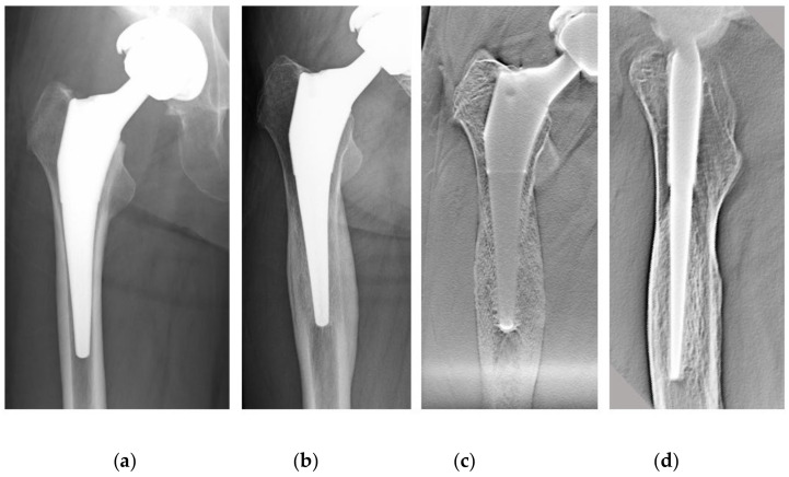
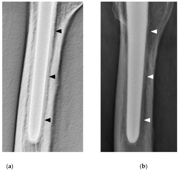
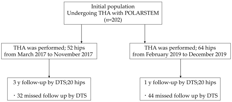
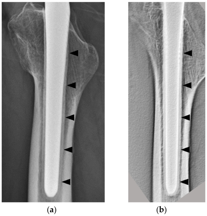
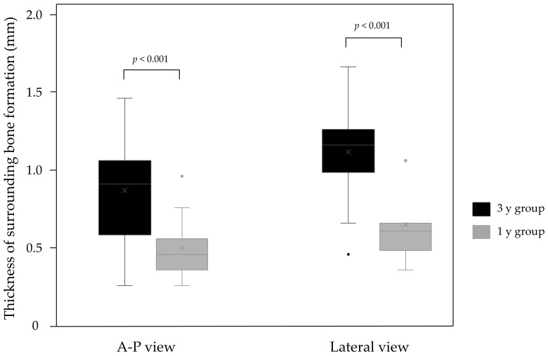
Digital tomosynthesis (DTS) is a new imaging technique derived from radiography, and its usefulness has been gradually reported in the field of orthopedic diagnosis in recent years. A fully hydroxyapatite (HA)-coated stem, which is used for total hip arthroplasty (THA), is a type of cementless stem that has been widely used recently and reported to have good results. However, stem loosening on plain radiographs is difficult to determine in some cases due to cancellous condensation around the stem. In this retrospective cross-sectional study, we compared the results of plain radiography versus DTS to evaluate the imaging findings after THA using a fully HA-coated stem. Twenty joints each in the 3 y and 1 y postoperative groups underwent plain radiography and DTS. On DTS, bone formation around the stem was confirmed in all cases; however, this formation was not reproducible on plain radiography, and there were cases in which the reaction could not be confirmed or cases with cancellous condensation resembling reactive lines. This reaction was not reproducible on plain radiographs, and in some cases, the reaction could not be confirmed, or there were cases with cancellous condensation that resembled reactive lines. Therefore, DTS was useful in the diagnosis of bone formation around the implant.
Keywords: POLARSTEM; digital tomosynthesis; fully hydroxyapatite-coated stem; surrounding bone formation.
Conflict of interest statement
The authors declare no conflict of interest.
Figures
References
-
- Ben-Shlomo Y., Blom A., Boulton C., Brittain R., Clark E., Craig R., Dawson-Bowling S., Deere K., Esler C., Espinoza O., et al. The National Joint Registry 17th Annual Report 2020. National Joint Registry; London, UK: 2020. National Joint Registry Annual Reports; pp. 41–119. - PubMed
LinkOut - more resources
Full Text Sources
