

Low-Dose PET Imaging of Tumors in Lung and Liver Regions Using Internal Motion Estimation
- PMID: 34829485
- PMCID: PMC8625002
- DOI: 10.3390/diagnostics11112138
Low-Dose PET Imaging of Tumors in Lung and Liver Regions Using Internal Motion Estimation
Abstract
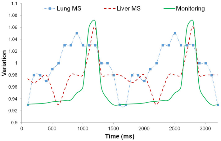
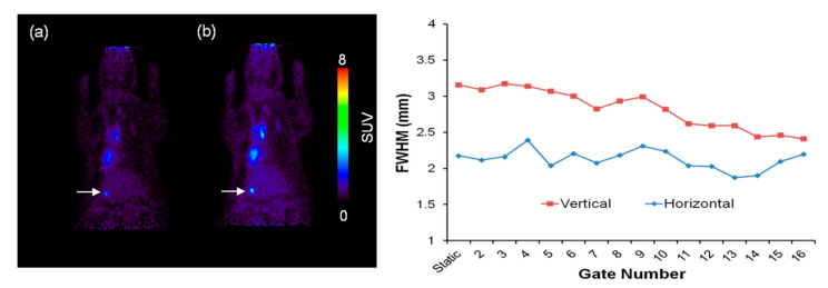
Motion estimation and compensation are necessary for improvement of tumor quantification analysis in positron emission tomography (PET) images. The aim of this study was to propose adaptive PET imaging with internal motion estimation and correction using regional artificial evaluation of tumors injected with low-dose and high-dose radiopharmaceuticals. In order to assess internal motion, molecular sieves imitating tumors were loaded with 18F and inserted into the lung and liver regions in rats. All models were classified into two groups, based on the injected radiopharmaceutical activity, to compare the effect of tumor intensity. The PET study was performed with injection of F-18 fluorodeoxyglucose (18F-FDG). Respiratory gating was carried out by external trigger device. Count, signal to noise ratio (SNR), contrast and full width at half maximum (FWHM) were measured in artificial tumors in gated images. Motion correction was executed by affine transformation with estimated internal motion data. Monitoring data were different from estimated motion. Contrast in the low-activity group was 3.57, 4.08 and 6.19, while in the high-activity group it was 10.01, 8.36 and 6.97 for static, 4 bin and 8 bin images, respectively. The results of the lung target in 4 bin and the liver target in 8 bin showed improvement in FWHM and contrast with sufficient SNR. After motion correction, FWHM was improved in both regions (lung: 24.56%, liver: 10.77%). Moreover, with the low dose of radiopharmaceuticals the PET image visualized specific accumulated radiopharmaceutical areas in the liver. Therefore, low activity in PET images should undergo motion correction before quantification analysis using PET data. We could improve quantitative tumor evaluation by considering organ region and tumor intensity.
Keywords: animal model; imaging; radioisotope; rat; respiratory organ.
Conflict of interest statement
The authors declare no conflict of interest.
Figures


References
-
- Lu Y., Fontaine K., Mulnix T., Onofrey J.A., Ren S., Panin V., Jones J., Casey M.E., Barnett R., Kench P., et al. Respiratory motion compensation for PET/CT with motion information derived from matched attenuation-corrected gated PET data. J. Nucl. Med. 2018;59:1480–1486. doi: 10.2967/jnumed.117.203000. - DOI - PMC - PubMed
-
- Mizuno H., Saito O., Tajiri M., Kimura T., Kuroiwa D., Shirai T., Inaniwa T., Fukahori M., Miki K., Fukuda S. Commissioning of a respiratory gating system involving a pressure sensor in carbon-ion scanning radiotherapy. J. Appl. Clin. Med. Phys. 2019;20:37–42. doi: 10.1002/acm2.12463. - DOI - PMC - PubMed
-
- Hurley S., Spangler-Bickell M., Deller T., Bradshaw T., Jansen F., McMillan A. Data-driven rigid motion correction of PET brain images using list mode reconstruction. J. Nucl. Med. 2019;60((Suppl. 1)):1358
Grants and funding
LinkOut - more resources
Full Text Sources
